1O23

CRYSTAL STRUCTURE OF LACTOSE SYNTHASE IN THE PRESENCE OF UDP-GLUCOSE


Experimental Data Snapshot

  • Method: X-RAY DIFFRACTION
  • Resolution: 2.32 Å
  • R-Value Free: 0.265 
  • R-Value Work: 0.201 
  • R-Value Observed: 0.201 

wwPDB Validation   3D Report Full Report


Ligand Structure Quality Assessment 


This is version 1.4 of the entry. See complete history


Literature

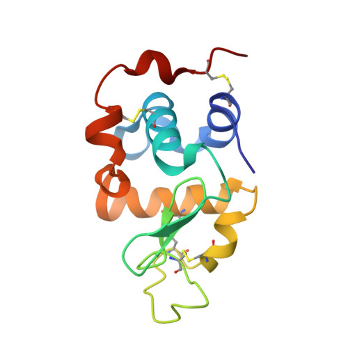
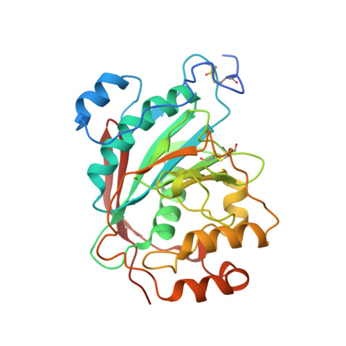
Alpha-Lactalbumin (La) Stimulates Milk Beta-1,4-Galactosyltransferase I (Beta 4Gal-T1) to Transfer Glucose from Udp-Glucose to N-Acetylglucosamine. Crystal Structure of Beta 4Gal-T1 X La Complex with Udp-Glc.

Ramakrishnan, B.Shah, P.S.Qasba, P.K.

(2001) J Biol Chem 276: 37665-37671

  • DOI: https://doi.org/10.1074/jbc.M102458200
  • Primary Citation of Related Structures:  
    1NWG, 1O23

  • PubMed Abstract: 

    beta-1,4-Galactosyltransferase 1 (Gal-T1) transfers galactose (Gal) from UDP-Gal to N-acetylglucosamine (GlcNAc), which constitutes its normal galactosyltransferase (Gal-T) activity. In the presence of alpha-lactalbumin (LA), it transfers Gal to Glc, which is its lactose synthase (LS) activity. It also transfers glucose (Glc) from UDP-Glc to GlcNAc, constituting the glucosyltransferase (Glc-T) activity, albeit at an efficiency of only 0.3-0.4% of Gal-T activity. In the present study, we show that LA increases this activity almost 30-fold. It also enhances the Glc-T activity toward various N-acyl substituted glucosamine acceptors. Steady state kinetic studies of Glc-T reaction show that the K(m) for the donor and acceptor substrates are high in the absence of LA. In the presence of LA, the K(m) for the acceptor substrate is reduced 30-fold, whereas for UDP-Glc it is reduced only 5-fold. In order to understand this property, we have determined the crystal structures of the Gal-T1.LA complex with UDP-Glc x Mn(2+) and with N-butanoyl-glucosamine (N-butanoyl-GlcN), a preferred sugar acceptor in the Glc-T activity. The crystal structures reveal that although the binding of UDP-Glc is quite similar to UDP-Gal, there are few significant differences observed in the hydrogen bonding interactions between UDP-Glc and Gal-T1. Based on the present kinetic and crystal structural studies, a possible explanation for the role of LA in the Glc-T activity has been proposed.


  • Organizational Affiliation

    Structural Glycobiology Section, Laboratory of Experimental and Computational Biology, Center for Cancer Research, NCI-Frederick, National Institutes of Health, Frederick, Maryland 21702, USA.


Macromolecules
Find similar proteins by:  (by identity cutoff)  |  3D Structure
Entity ID: 1
MoleculeChains Sequence LengthOrganismDetailsImage
ALPHA-LACTALBUMIN
A, C
123Mus musculusMutation(s): 0 
Gene Names: LALBA
UniProt
Find proteins for P29752 (Mus musculus)
Explore P29752 
Go to UniProtKB:  P29752
Entity Groups  
Sequence Clusters30% Identity50% Identity70% Identity90% Identity95% Identity100% Identity
UniProt GroupP29752
Sequence Annotations
Expand
  • Reference Sequence
Find similar proteins by:  (by identity cutoff)  |  3D Structure
Entity ID: 2
MoleculeChains Sequence LengthOrganismDetailsImage
BETA-1,4-GALACTOSYLTRANSFERASE
B, D
286Bos taurusMutation(s): 0 
Gene Names: BETA14-GALACTOSYLTRANSFERASE
EC: 2.4.1.90
UniProt
Find proteins for P08037 (Bos taurus)
Explore P08037 
Go to UniProtKB:  P08037
Entity Groups  
Sequence Clusters30% Identity50% Identity70% Identity90% Identity95% Identity100% Identity
UniProt GroupP08037
Sequence Annotations
Expand
  • Reference Sequence
Small Molecules
Ligands 6 Unique
IDChains Name / Formula / InChI Key2D Diagram3D Interactions
UPG
Query on UPG

Download Ideal Coordinates CCD File 
I [auth B],
N [auth D]
URIDINE-5'-DIPHOSPHATE-GLUCOSE
C15 H24 N2 O17 P2
HSCJRCZFDFQWRP-JZMIEXBBSA-N
UDP
Query on UDP

Download Ideal Coordinates CCD File 
J [auth B],
O [auth D]
URIDINE-5'-DIPHOSPHATE
C9 H14 N2 O12 P2
XCCTYIAWTASOJW-XVFCMESISA-N
MES
Query on MES

Download Ideal Coordinates CCD File 
G [auth A]2-(N-MORPHOLINO)-ETHANESULFONIC ACID
C6 H13 N O4 S
SXGZJKUKBWWHRA-UHFFFAOYSA-N
PG4
Query on PG4

Download Ideal Coordinates CCD File 
F [auth A],
L [auth C]
TETRAETHYLENE GLYCOL
C8 H18 O5
UWHCKJMYHZGTIT-UHFFFAOYSA-N
MN
Query on MN

Download Ideal Coordinates CCD File 
H [auth B],
M [auth D]
MANGANESE (II) ION
Mn
WAEMQWOKJMHJLA-UHFFFAOYSA-N
CA
Query on CA

Download Ideal Coordinates CCD File 
E [auth A],
K [auth C]
CALCIUM ION
Ca
BHPQYMZQTOCNFJ-UHFFFAOYSA-N
Experimental Data & Validation

Experimental Data

  • Method: X-RAY DIFFRACTION
  • Resolution: 2.32 Å
  • R-Value Free: 0.265 
  • R-Value Work: 0.201 
  • R-Value Observed: 0.201 
  • Space Group: P 1 21 1
Unit Cell:
Length ( Å )Angle ( ˚ )
a = 55.545α = 90
b = 99.379β = 104.09
c = 102.568γ = 90
Software Package:
Software NamePurpose
DENZOdata reduction
SCALEPACKdata scaling
AMoREphasing
CNSrefinement

Structure Validation

View Full Validation Report


Ligand Structure Quality Assessment 


Entry History 

Deposition Data

Revision History  (Full details and data files)

  • Version 1.0: 2003-02-25
    Type: Initial release
  • Version 1.1: 2008-04-26
    Changes: Version format compliance
  • Version 1.2: 2011-07-13
    Changes: Version format compliance
  • Version 1.3: 2013-05-08
    Changes: Non-polymer description
  • Version 1.4: 2023-12-27
    Changes: Data collection, Database references, Derived calculations