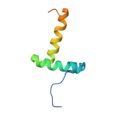
Solution structure of Switch Arc, a mutant with 3(10) helices replacing a wild-type beta-ribbon
Cordes, M.H., Walsh, N.P., McKnight, C.J., Sauer, R.T.(2003) J Mol Biol 326: 899-909
- PubMed: 12581649
- DOI: https://doi.org/10.1016/s0022-2836(02)01425-0
- Primary Citation of Related Structures:
1NLA - PubMed Abstract:
Adjacent N11L and L12N mutations in the antiparallel beta-ribbon of Arc repressor result in dramatic changes in local structure in which each beta-strand is replaced by a right-handed helix. The full solution structure of this "switch" Arc mutant shows that irregular 3(10) helices compose the new secondary structure. This structural metamorphosis conserves the number of main-chain and side-chain to main-chain hydrogen bonds and the number of fully buried core residues. Apart from a slight widening of the interhelical angle between alpha-helices A and B and changes in side-chain conformation of a few core residues in Arc, no large-scale structural adjustments in the remainder of the protein are necessary to accommodate the ribbon-to-helix change. Nevertheless, some changes in hydrogen-exchange rates are observed, even in regions that have very similar structures in the two proteins. The surface of switch Arc is packed poorly compared to wild-type, leading to approximately 1000A(2) of additional solvent-accessible surface area, and the N termini of the 3(10) helices make unfavorable head-to-head electrostatic interactions. These structural features account for the positive m value and salt dependence of the ribbon-to-helix transition in Arc-N11L, a variant that can adopt either the mutant or wild-type structures. The tertiary fold is capped in different ways in switch and wild-type Arc, showing how stepwise evolutionary transformations can arise through small changes in amino acid sequence.
Organizational Affiliation:
Department of Biology, Massachusetts Institute of Technology, Cambridge 02139, MA, USA. cordes@email.arizona.edu