1NKH

Crystal structure of Lactose synthase complex with UDP and Manganese


Experimental Data Snapshot

  • Method: X-RAY DIFFRACTION
  • Resolution: 2.00 Å
  • R-Value Free: 0.251 
  • R-Value Work: 0.196 
  • R-Value Observed: 0.196 

wwPDB Validation   3D Report Full Report


Ligand Structure Quality Assessment 


This is version 1.2 of the entry. See complete history


Literature

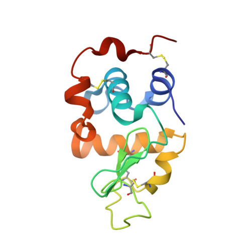
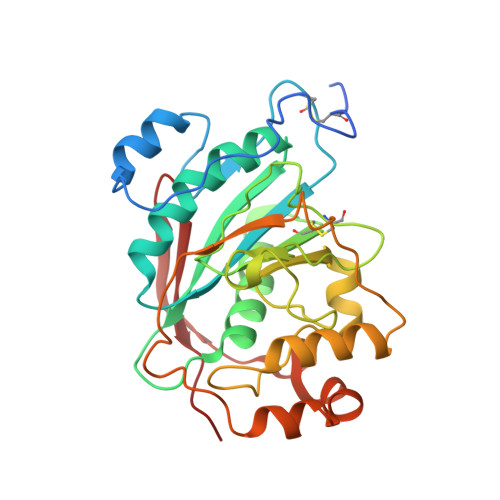
Crystal structure of lactose synthase reveals a large conformational change in its catalytic component, the beta-1,4-galactosyltransferase

Ramakrishnan, B.Qasba, P.K.

(2001) J Mol Biol 310: 205-218

  • DOI: https://doi.org/10.1006/jmbi.2001.4757
  • Primary Citation of Related Structures:  
    1NHE, 1NKH

  • PubMed Abstract: 

    The lactose synthase (LS) enzyme is a 1:1 complex of a catalytic component, beta1,4-galactosyltransferse (beta4Gal-T1) and a regulatory component, alpha-lactalbumin (LA), a mammary gland-specific protein. LA promotes the binding of glucose (Glc) to beta4Gal-T1, thereby altering its sugar acceptor specificity from N-acetylglucosamine (GlcNAc) to glucose, which enables LS to synthesize lactose, the major carbohydrate component of milk. The crystal structures of LS bound with various substrates were solved at 2 A resolution. These structures reveal that upon substrate binding to beta4Gal-T1, a large conformational change occurs in the region comprising residues 345 to 365. This repositions His347 in such a way that it can participate in the coordination of a metal ion, and creates a sugar and LA-binding site. At the sugar-acceptor binding site, a hydrophobic N-acetyl group-binding pocket is found, formed by residues Arg359, Phe360 and Ile363. In the Glc-bound structure, this hydrophobic pocket is absent. For the binding of Glc to LS, a reorientation of the Arg359 side-chain occurs, which blocks the hydrophobic pocket and maximizes the interactions with the Glc molecule. Thus, the role of LA is to hold Glc by hydrogen bonding with the O-1 hydroxyl group in the acceptor-binding site on beta4Gal-T1, while the N-acetyl group-binding pocket in beta4Gal-T1 adjusts to maximize the interactions with the Glc molecule. This study provides details of a structural basis for the partially ordered kinetic mechanism proposed for lactose synthase.


  • Organizational Affiliation

    Structural Glycobiology Section, Intramural Research Support Program-SAIC, Laboratory of Experimental and Computational Biology, CCR, NCI, Frederick, MD 21702, USA.


Macromolecules
Find similar proteins by:  (by identity cutoff)  |  3D Structure
Entity ID: 1
MoleculeChains Sequence LengthOrganismDetailsImage
ALPHA-LACTALBUMIN
A, C
123Mus musculusMutation(s): 0 
Gene Names: alpha-lactalbumin
UniProt
Find proteins for P29752 (Mus musculus)
Explore P29752 
Go to UniProtKB:  P29752
Entity Groups  
Sequence Clusters30% Identity50% Identity70% Identity90% Identity95% Identity100% Identity
UniProt GroupP29752
Sequence Annotations
Expand
  • Reference Sequence
Find similar proteins by:  (by identity cutoff)  |  3D Structure
Entity ID: 2
MoleculeChains Sequence LengthOrganismDetailsImage
BETA-1,4-GALACTOSYLTRANSFERASE
B, D
286Bos taurusMutation(s): 0 
Gene Names: beta14-galactosyltransferase
EC: 2.4.1.22 (PDB Primary Data), 2.4.1.90 (PDB Primary Data), 2.4.1.38 (PDB Primary Data)
UniProt
Find proteins for P08037 (Bos taurus)
Explore P08037 
Go to UniProtKB:  P08037
Entity Groups  
Sequence Clusters30% Identity50% Identity70% Identity90% Identity95% Identity100% Identity
UniProt GroupP08037
Sequence Annotations
Expand
  • Reference Sequence
Small Molecules
Ligands 5 Unique
IDChains Name / Formula / InChI Key2D Diagram3D Interactions
UDP
Query on UDP

Download Ideal Coordinates CCD File 
I [auth B],
J [auth B],
Q [auth D],
R [auth D]
URIDINE-5'-DIPHOSPHATE
C9 H14 N2 O12 P2
XCCTYIAWTASOJW-XVFCMESISA-N
PG4
Query on PG4

Download Ideal Coordinates CCD File 
G [auth A],
K [auth B],
L [auth B],
N [auth C],
S [auth D]
TETRAETHYLENE GLYCOL
C8 H18 O5
UWHCKJMYHZGTIT-UHFFFAOYSA-N
SO4
Query on SO4

Download Ideal Coordinates CCD File 
F [auth A],
P [auth D]
SULFATE ION
O4 S
QAOWNCQODCNURD-UHFFFAOYSA-L
MN
Query on MN

Download Ideal Coordinates CCD File 
H [auth B],
O [auth D]
MANGANESE (II) ION
Mn
WAEMQWOKJMHJLA-UHFFFAOYSA-N
CA
Query on CA

Download Ideal Coordinates CCD File 
E [auth A],
M [auth C]
CALCIUM ION
Ca
BHPQYMZQTOCNFJ-UHFFFAOYSA-N
Experimental Data & Validation

Experimental Data

  • Method: X-RAY DIFFRACTION
  • Resolution: 2.00 Å
  • R-Value Free: 0.251 
  • R-Value Work: 0.196 
  • R-Value Observed: 0.196 
  • Space Group: P 1 21 1
Unit Cell:
Length ( Å )Angle ( ˚ )
a = 55.469α = 90
b = 99.145β = 104.14
c = 102.261γ = 90
Software Package:
Software NamePurpose
DENZOdata reduction
SCALEPACKdata scaling
AMoREphasing
CNSrefinement

Structure Validation

View Full Validation Report


Ligand Structure Quality Assessment 


Entry History 

Deposition Data

Revision History  (Full details and data files)

  • Version 1.0: 2003-01-14
    Type: Initial release
  • Version 1.1: 2008-04-29
    Changes: Version format compliance
  • Version 1.2: 2011-07-13
    Changes: Version format compliance