1N52

Cap Binding Complex


Experimental Data Snapshot

  • Method: X-RAY DIFFRACTION
  • Resolution: 2.11 Å
  • R-Value Free: 0.246 
  • R-Value Work: 0.223 

wwPDB Validation   3D Report Full Report


Ligand Structure Quality Assessment 


This is version 1.3 of the entry. See complete history


Literature

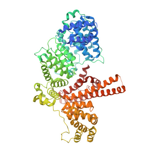
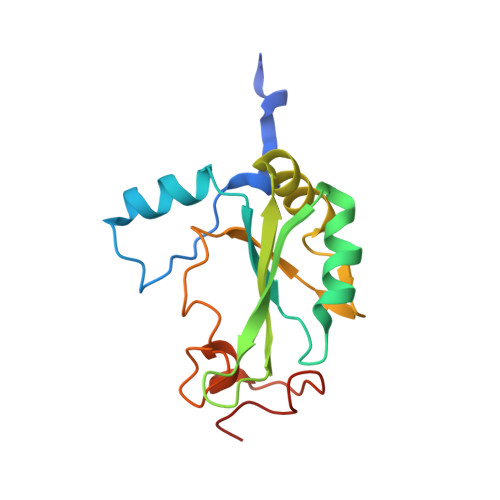
Structural basis of m7GpppG binding to the nuclear cap-binding protein complex.

Calero, G.Wilson, K.Ly, T.Rios-Steiner, J.Clardy, J.Cerione, R.

(2002) Nat Struct Biol 9: 912-917

  • DOI: https://doi.org/10.1038/nsb874
  • Primary Citation of Related Structures:  
    1N52, 1N54

  • PubMed Abstract: 

    The 7-methyl guanosine cap structure of RNA is essential for key aspects of RNA processing, including pre-mRNA splicing, 3' end formation, U snRNA transport, nonsense-mediated decay and translation. Two cap-binding proteins mediate these effects: cytosolic eIF-4E and nuclear cap-binding protein complex (CBC). The latter consists of a CBP20 subunit, which binds the cap, and a CBP80 subunit, which ensures high-affinity cap binding. Here we report the 2.1 A resolution structure of human CBC with the cap analog m7GpppG, as well as the structure of unliganded CBC. Comparisons between these structures indicate that the cap induces substantial conformational changes within the N-terminal loop of CBP20, enabling Tyr 20 to join Tyr 43 in pi-pi stacking interactions with the methylated guanosine base. CBP80 stabilizes the movement of the N-terminal loop of CBP20 and locks the CBC into a high affinity cap-binding state. The structure for the CBC bound to m7GpppG highlights interesting similarities and differences between CBC and eIF-4E, and provides insights into the regulatory mechanisms used by growth factors and other extracellular stimuli to influence the cap-binding state of the CBC.


  • Organizational Affiliation

    Department of Chemistry and Chemical Biology, Baker Laboratory, Cornell University, Ithaca, New York 14853, USA.


Macromolecules
Find similar proteins by:  (by identity cutoff)  |  3D Structure
Entity ID: 1
MoleculeChains Sequence LengthOrganismDetailsImage
80 kDa nuclear cap binding protein790Homo sapiensMutation(s): 0 
Gene Names: CBP80
UniProt & NIH Common Fund Data Resources
Find proteins for Q09161 (Homo sapiens)
Explore Q09161 
Go to UniProtKB:  Q09161
PHAROS:  Q09161
GTEx:  ENSG00000136937 
Entity Groups  
Sequence Clusters30% Identity50% Identity70% Identity90% Identity95% Identity100% Identity
UniProt GroupQ09161
Sequence Annotations
Expand
  • Reference Sequence
Find similar proteins by:  (by identity cutoff)  |  3D Structure
Entity ID: 2
MoleculeChains Sequence LengthOrganismDetailsImage
20 kDa nuclear cap binding protein156Homo sapiensMutation(s): 0 
Gene Names: CBP20
UniProt & NIH Common Fund Data Resources
Find proteins for P52298 (Homo sapiens)
Explore P52298 
Go to UniProtKB:  P52298
PHAROS:  P52298
GTEx:  ENSG00000114503 
Entity Groups  
Sequence Clusters30% Identity50% Identity70% Identity90% Identity95% Identity100% Identity
UniProt GroupP52298
Sequence Annotations
Expand
  • Reference Sequence
Small Molecules
Ligands 4 Unique
IDChains Name / Formula / InChI Key2D Diagram3D Interactions
GTG
Query on GTG

Download Ideal Coordinates CCD File 
P [auth B]7-METHYL-GUANOSINE-5'-TRIPHOSPHATE-5'-GUANOSINE
C21 H30 N10 O18 P3
FHHZHGZBHYYWTG-INFSMZHSSA-O
PG4
Query on PG4

Download Ideal Coordinates CCD File 
D [auth A]TETRAETHYLENE GLYCOL
C8 H18 O5
UWHCKJMYHZGTIT-UHFFFAOYSA-N
GOL
Query on GOL

Download Ideal Coordinates CCD File 
E [auth A]
F [auth A]
G [auth A]
H [auth A]
I [auth A]
E [auth A],
F [auth A],
G [auth A],
H [auth A],
I [auth A],
J [auth A],
K [auth A],
L [auth A],
M [auth A],
N [auth A],
O [auth A],
Q [auth B],
R [auth B],
S [auth B],
T [auth B],
U [auth B]
GLYCEROL
C3 H8 O3
PEDCQBHIVMGVHV-UHFFFAOYSA-N
MG
Query on MG

Download Ideal Coordinates CCD File 
C [auth A]MAGNESIUM ION
Mg
JLVVSXFLKOJNIY-UHFFFAOYSA-N
Experimental Data & Validation

Experimental Data

  • Method: X-RAY DIFFRACTION
  • Resolution: 2.11 Å
  • R-Value Free: 0.246 
  • R-Value Work: 0.223 
  • Space Group: P 32 2 1
Unit Cell:
Length ( Å )Angle ( ˚ )
a = 111.681α = 90
b = 111.681β = 90
c = 177.588γ = 120
Software Package:
Software NamePurpose
CNSrefinement
DENZOdata reduction
SCALEPACKdata scaling
MOLREPphasing

Structure Validation

View Full Validation Report


Ligand Structure Quality Assessment 


Entry History 

Deposition Data

Revision History  (Full details and data files)

  • Version 1.0: 2003-02-18
    Type: Initial release
  • Version 1.1: 2008-04-28
    Changes: Version format compliance
  • Version 1.2: 2011-07-13
    Changes: Non-polymer description, Version format compliance
  • Version 1.3: 2024-02-14
    Changes: Data collection, Database references, Derived calculations, Refinement description