1N1F

Crystal Structure of Human Interleukin-19


Experimental Data Snapshot

  • Method: X-RAY DIFFRACTION
  • Resolution: 1.95 Å
  • R-Value Free: 0.243 
  • R-Value Work: 0.156 
  • R-Value Observed: 0.156 

wwPDB Validation   3D Report Full Report


Ligand Structure Quality Assessment 


This is version 1.3 of the entry. See complete history


Literature

Crystal structure of interleukin-19 defines a new subfamily of helical cytokines

Chang, C.Magracheva, E.Kozlov, S.Fong, S.Tobin, G.Kotenko, S.Wlodawer, A.Zdanov, A.

(2003) J Biol Chem 278: 3308-3313

  • DOI: https://doi.org/10.1074/jbc.M208602200
  • Primary Citation of Related Structures:  
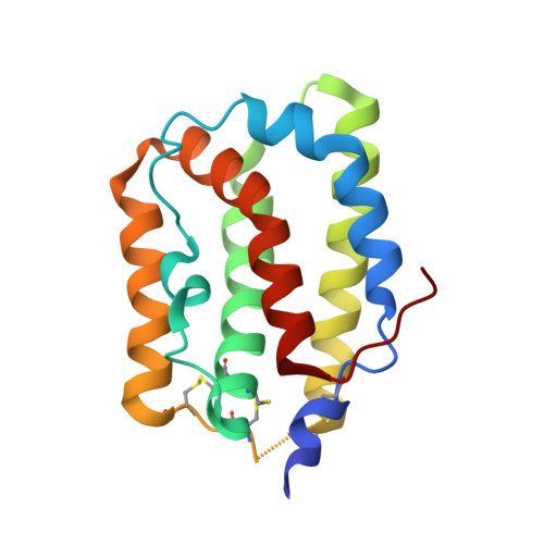
    1N1F

  • PubMed Abstract: 

    Interleukin-19 (IL-19) is a novel cytokine that was initially identified during a sequence data base search aimed at finding potential IL-10 homologs. IL-19 shares a receptor complex with IL-20, indicating that the biological activities of these two cytokines overlap and that both may play an important role in regulating development and proper functioning of the skin. We determined the crystal structure of human recombinant IL-19 and refined it at 1.95-A resolution to an R-factor of 0.157. Unlike IL-10, which forms an intercalated dimer, the molecule of IL-19 is a monomer made of seven amphipathic helices, A-G, creating a unique helical bundle. On the basis of the observed structure, we propose that IL-19, IL-20, and other putative members of the proposed IL-10 family together form a distinct subfamily of helical cytokines.


  • Organizational Affiliation

    Protein Structure Section, Macromolecular Crystallography Laboratory, Center for Cancer Research, National Cancer Institute-Frederick, National Institutes of Health, Maryland 21702-1201, USA.


Macromolecules
Find similar proteins by:  (by identity cutoff)  |  3D Structure
Entity ID: 1
MoleculeChains Sequence LengthOrganismDetailsImage
interleukin-19159Homo sapiensMutation(s): 0 
UniProt & NIH Common Fund Data Resources
Find proteins for Q9UHD0 (Homo sapiens)
Explore Q9UHD0 
Go to UniProtKB:  Q9UHD0
PHAROS:  Q9UHD0
GTEx:  ENSG00000142224 
Entity Groups  
Sequence Clusters30% Identity50% Identity70% Identity90% Identity95% Identity100% Identity
UniProt GroupQ9UHD0
Sequence Annotations
Expand
  • Reference Sequence
Small Molecules
Ligands 1 Unique
IDChains Name / Formula / InChI Key2D Diagram3D Interactions
NAG
Query on NAG

Download Ideal Coordinates CCD File 
B [auth A]2-acetamido-2-deoxy-beta-D-glucopyranose
C8 H15 N O6
OVRNDRQMDRJTHS-FMDGEEDCSA-N
Experimental Data & Validation

Experimental Data

  • Method: X-RAY DIFFRACTION
  • Resolution: 1.95 Å
  • R-Value Free: 0.243 
  • R-Value Work: 0.156 
  • R-Value Observed: 0.156 
  • Space Group: P 21 21 21
Unit Cell:
Length ( Å )Angle ( ˚ )
a = 30.657α = 90
b = 52.974β = 90
c = 93.474γ = 90
Software Package:
Software NamePurpose
DENZOdata reduction
SCALEPACKdata scaling
SOLVEphasing
CNSrefinement

Structure Validation

View Full Validation Report


Ligand Structure Quality Assessment 


Entry History 

Deposition Data

Revision History  (Full details and data files)

  • Version 1.0: 2003-02-04
    Type: Initial release
  • Version 1.1: 2008-04-28
    Changes: Version format compliance
  • Version 1.2: 2011-07-13
    Changes: Non-polymer description, Version format compliance
  • Version 1.3: 2020-07-29
    Type: Remediation
    Reason: Carbohydrate remediation
    Changes: Data collection, Derived calculations, Structure summary