1MT6

Structure of histone H3 K4-specific methyltransferase SET7/9 with AdoHcy


Experimental Data Snapshot

  • Method: X-RAY DIFFRACTION
  • Resolution: 2.20 Å
  • R-Value Free: 0.263 
  • R-Value Work: 0.226 
  • R-Value Observed: 0.226 

wwPDB Validation   3D Report Full Report


Ligand Structure Quality Assessment 


This is version 1.3 of the entry. See complete history


Literature

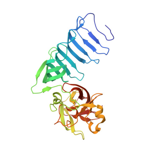
The active site of the SET domain is constructed on a knot

Jacobs, S.A.Harp, J.M.Devarakonda, S.Kim, Y.Rastinejad, F.Khorasanizadeh, S.

(2002) Nat Struct Biol 9: 833-838

  • DOI: https://doi.org/10.1038/nsb861
  • Primary Citation of Related Structures:  
    1MT6, 1MUF

  • PubMed Abstract: 

    The SET domain contains the catalytic center of lysine methyltransferases that target the N-terminal tails of histones and regulate chromatin function. Here we report the structure of the SET7/9 protein in the absence and presence of its cofactor product, S-adenosyl-L-homocysteine (AdoHcy). A knot within the SET domain helps form the methyltransferase active site, where AdoHcy binds and lysine methylation is likely to occur. A structure-guided comparison of sequences within the SET protein family suggests that the knot substructure and active site environment are conserved features of the SET domain.


  • Organizational Affiliation

    Department of Biochemistry and Molecular Genetics, University of Virginia Health System, Charlottesville, Virginia 22908, USA.


Macromolecules
Find similar proteins by:  (by identity cutoff)  |  3D Structure
Entity ID: 1
MoleculeChains Sequence LengthOrganismDetailsImage
SET9280Homo sapiensMutation(s): 0 
EC: 2.1.1.43
UniProt & NIH Common Fund Data Resources
Find proteins for Q8WTS6 (Homo sapiens)
Explore Q8WTS6 
Go to UniProtKB:  Q8WTS6
PHAROS:  Q8WTS6
GTEx:  ENSG00000145391 
Entity Groups  
Sequence Clusters30% Identity50% Identity70% Identity90% Identity95% Identity100% Identity
UniProt GroupQ8WTS6
Sequence Annotations
Expand
  • Reference Sequence
Small Molecules
Ligands 1 Unique
IDChains Name / Formula / InChI Key2D Diagram3D Interactions
SAH
Query on SAH

Download Ideal Coordinates CCD File 
B [auth A]S-ADENOSYL-L-HOMOCYSTEINE
C14 H20 N6 O5 S
ZJUKTBDSGOFHSH-WFMPWKQPSA-N
Binding Affinity Annotations 
IDSourceBinding Affinity
SAH BindingDB:  1MT6 IC50: min: 3.00e+4, max: 4.70e+4 (nM) from 2 assay(s)
Experimental Data & Validation

Experimental Data

  • Method: X-RAY DIFFRACTION
  • Resolution: 2.20 Å
  • R-Value Free: 0.263 
  • R-Value Work: 0.226 
  • R-Value Observed: 0.226 
  • Space Group: I 2 2 2
Unit Cell:
Length ( Å )Angle ( ˚ )
a = 70.347α = 90
b = 76.038β = 90
c = 166.284γ = 90
Software Package:
Software NamePurpose
HKL-2000data collection
SCALEPACKdata scaling
CNSrefinement
HKL-2000data reduction
CNSphasing

Structure Validation

View Full Validation Report


Ligand Structure Quality Assessment 


Entry History 

Deposition Data

Revision History  (Full details and data files)

  • Version 1.0: 2002-11-06
    Type: Initial release
  • Version 1.1: 2008-04-28
    Changes: Version format compliance
  • Version 1.2: 2011-07-13
    Changes: Version format compliance
  • Version 1.3: 2024-02-14
    Changes: Data collection, Database references, Derived calculations