1MOH

FERRIC MONOMERIC HEMOGLOBIN I (HB I)


Experimental Data Snapshot

  • Method: X-RAY DIFFRACTION
  • Resolution: 1.90 Å

wwPDB Validation   3D Report Full Report


This is version 1.3 of the entry. See complete history


Literature

Structural bases for sulfide recognition in Lucina pectinata hemoglobin I.

Rizzi, M.Wittenberg, J.B.Coda, A.Ascenzi, P.Bolognesi, M.

(1996) J Mol Biol 258: 1-5

  • DOI: https://doi.org/10.1006/jmbi.1996.0228
  • Primary Citation of Related Structures:  
    1MOH

  • PubMed Abstract: 

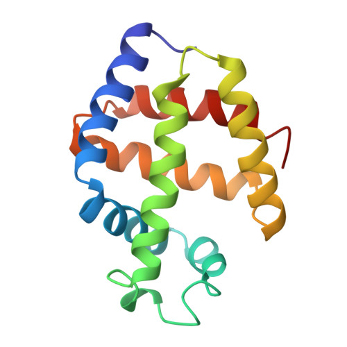
    The X-ray crystal structure of the sulfide derivative of ferric Lucina pectinata hemoglobin component I (HbI) has been determined at 1.9 A resolution (R-factor 0.186). The heme pocket structural organization of HbI is in keeping with its ligand binding properties. The fast sulfide association rate constant can be related to the presence of Gln(64)E7, as the heme distal residue, together with the protein structural properties in the CD-E distal region. Moreover, the very high sulfide affinity for HbI is reflected by the exceptionally slow ligand dissociation rate. The stabilization of the heme-bound sulfide molecule is achieved through hydrogen bonding to Gln(64)E7, as well as by finely tuned aromatic-electrostatic interactions with the clustered residues Phe(29)B10, Phe(43)CD1 and Phe(68)E11. Such a peculiar arrangement of phenylalanyl residues at the distal ligand binding site has not been observed before in the globin family, and is unique to HbI, a protein functionally devoted to sulfide transport.


  • Organizational Affiliation

    Dipartimento di Genetica e Microbiologia, Università di Pavia, Italy.


Macromolecules
Find similar proteins by:  (by identity cutoff)  |  3D Structure
Entity ID: 1
MoleculeChains Sequence LengthOrganismDetailsImage
MONOMERIC HEMOGLOBIN I142Phacoides pectinatusMutation(s): 0 
UniProt
Find proteins for P41260 (Phacoides pectinatus)
Explore P41260 
Go to UniProtKB:  P41260
Entity Groups  
Sequence Clusters30% Identity50% Identity70% Identity90% Identity95% Identity100% Identity
UniProt GroupP41260
Sequence Annotations
Expand
  • Reference Sequence
Small Molecules
Ligands 2 Unique
IDChains Name / Formula / InChI Key2D Diagram3D Interactions
HEM
Query on HEM

Download Ideal Coordinates CCD File 
B [auth A]PROTOPORPHYRIN IX CONTAINING FE
C34 H32 Fe N4 O4
KABFMIBPWCXCRK-RGGAHWMASA-L
H2S
Query on H2S

Download Ideal Coordinates CCD File 
C [auth A]HYDROSULFURIC ACID
H2 S
RWSOTUBLDIXVET-UHFFFAOYSA-N
Experimental Data & Validation

Experimental Data

  • Method: X-RAY DIFFRACTION
  • Resolution: 1.90 Å
  • Space Group: P 1 21 1
Unit Cell:
Length ( Å )Angle ( ˚ )
a = 50.55α = 90
b = 38.79β = 106.93
c = 42.51γ = 90
Software Package:
Software NamePurpose
TNTrefinement
MOSFLMdata reduction

Structure Validation

View Full Validation Report


Entry History 

Deposition Data

Revision History  (Full details and data files)

  • Version 1.0: 1996-08-01
    Type: Initial release
  • Version 1.1: 2008-03-24
    Changes: Version format compliance
  • Version 1.2: 2011-07-13
    Changes: Version format compliance
  • Version 1.3: 2024-02-14
    Changes: Data collection, Database references, Derived calculations, Other