1MGW

Crystal structure of RNase Sa3, cytotoxic microbial ribonuclease


Experimental Data Snapshot

  • Method: X-RAY DIFFRACTION
  • Resolution: 2.00 Å
  • R-Value Free: 0.212 
  • R-Value Work: 0.154 
  • R-Value Observed: 0.155 

wwPDB Validation   3D Report Full Report


This is version 1.3 of the entry. See complete history


Literature

Links X-ray Structure of Two Crystalline Forms of a Streptomycete Ribonuclease with Cytotoxic Activity

Sevcik, J.Urbanikova, L.Leland, P.A.Raines, R.T.

(2002) J Biol Chem 277: 47325-47330

  • DOI: https://doi.org/10.1074/jbc.M208425200
  • Primary Citation of Related Structures:  
    1MGR, 1MGW

  • PubMed Abstract: 

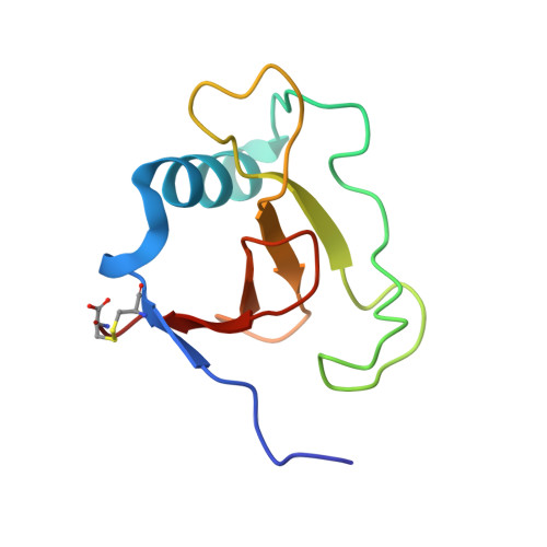
    Ribonuclease (RNase) Sa3 is secreted by the Gram-positive bacterium Streptomyces aureofaciens. The enzyme catalyzes the cleavage of RNA on the 3' side of guanosine residues. Here, x-ray diffraction analysis was used to determine the three-dimensional structure of two distinct crystalline forms of RNase Sa3 to a resolution of 2.0 and 1.7 A. These two structures are similar to each other as well as to that of a homolog, RNase Sa. All of the key active-site residues of RNase Sa (Asn(42), Glu(44), Glu(57), Arg(72), and His(88)) are located in the putative active site of RNase Sa3. Also herein, RNase Sa3 is shown to be toxic to human erythroleukemia cells in culture. Like onconase, which is an amphibian ribonuclease in Phase III clinical trials as a cancer chemotherapeutic, RNase Sa3 is not inhibited by the cytosolic ribonuclease inhibitor protein. Thus, a prokaryotic ribonuclease can be toxic to mammalian cells.


  • Organizational Affiliation

    Institute of Molecular Biology, Slovak Academy of Sciences, Dubravska cesta 21, 84251 Bratislava, Slovak Republic. umbisevc@savba.sk


Macromolecules
Find similar proteins by:  (by identity cutoff)  |  3D Structure
Entity ID: 1
MoleculeChains Sequence LengthOrganismDetailsImage
Guanyl-specific ribonuclease Sa399Kitasatospora aureofaciensMutation(s): 0 
EC: 3.1.27.3
UniProt
Find proteins for P30289 (Kitasatospora aureofaciens)
Explore P30289 
Go to UniProtKB:  P30289
Entity Groups  
Sequence Clusters30% Identity50% Identity70% Identity90% Identity95% Identity100% Identity
UniProt GroupP30289
Sequence Annotations
Expand
  • Reference Sequence
Small Molecules
Ligands 1 Unique
IDChains Name / Formula / InChI Key2D Diagram3D Interactions
LI
Query on LI

Download Ideal Coordinates CCD File 
B [auth A]LITHIUM ION
Li
HBBGRARXTFLTSG-UHFFFAOYSA-N
Experimental Data & Validation

Experimental Data

  • Method: X-RAY DIFFRACTION
  • Resolution: 2.00 Å
  • R-Value Free: 0.212 
  • R-Value Work: 0.154 
  • R-Value Observed: 0.155 
  • Space Group: P 31 2 1
Unit Cell:
Length ( Å )Angle ( ˚ )
a = 64.72α = 90
b = 64.72β = 90
c = 69.57γ = 120
Software Package:
Software NamePurpose
DENZOdata reduction
SCALEPACKdata scaling
AMoREphasing
REFMACrefinement

Structure Validation

View Full Validation Report


Entry History 

Deposition Data

Revision History  (Full details and data files)

  • Version 1.0: 2003-02-04
    Type: Initial release
  • Version 1.1: 2008-04-28
    Changes: Version format compliance
  • Version 1.2: 2011-07-13
    Changes: Version format compliance
  • Version 1.3: 2011-11-16
    Changes: Atomic model