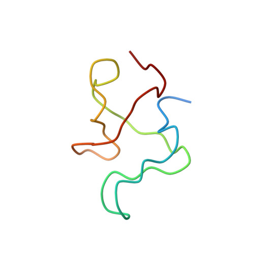
Structure of a protein determined by solid-state magic-angle-spinning NMR spectroscopy
Castellani, F., Van Rossum, B., Diehl, A., Schubert, M., Rehbein, K., Oschkinat, H.(2002) Nature 420: 98-102
- PubMed: 12422222
- DOI: https://doi.org/10.1038/nature01070
- Primary Citation of Related Structures:
1M8M - PubMed Abstract:
The determination of a representative set of protein structures is a chief aim in structural genomics. Solid-state NMR may have a crucial role in structural investigations of those proteins that do not easily form crystals or are not accessible to solution NMR, such as amyloid systems or membrane proteins. Here we present a protein structure determined by solid-state magic-angle-spinning (MAS) NMR. Almost complete (13)C and (15)N resonance assignments for a micro-crystalline preparation of the alpha-spectrin Src-homology 3 (SH3) domain formed the basis for the extraction of a set of distance restraints. These restraints were derived from proton-driven spin diffusion (PDSD) spectra of biosynthetically site-directed, labelled samples obtained from bacteria grown using [1,3-(13)C]glycerol or [2-(13)C]glycerol as carbon sources. This allowed the observation of long-range distance correlations up to approximately 7 A. The calculated global fold of the alpha-spectrin SH3 domain is based on 286 inter-residue (13)C-(13)C and six (15)N-(15)N restraints, all self-consistently obtained by solid-state MAS NMR. This MAS NMR procedure should be widely applicable to small membrane proteins that can be expressed in bacteria.
Organizational Affiliation:
Forschungsinstitut für Molekulare Pharmakologie, Robert-Rössle-Strasse 10, 13125 Berlin, Germany.