1M85

Structure of Proteus mirabilis catalase for the native form


Experimental Data Snapshot

  • Method: X-RAY DIFFRACTION
  • Resolution: 2.00 Å
  • R-Value Free: 0.218 
  • R-Value Work: 0.194 
  • R-Value Observed: 0.194 

wwPDB Validation   3D Report Full Report


Ligand Structure Quality Assessment 


This is version 1.3 of the entry. See complete history


Literature

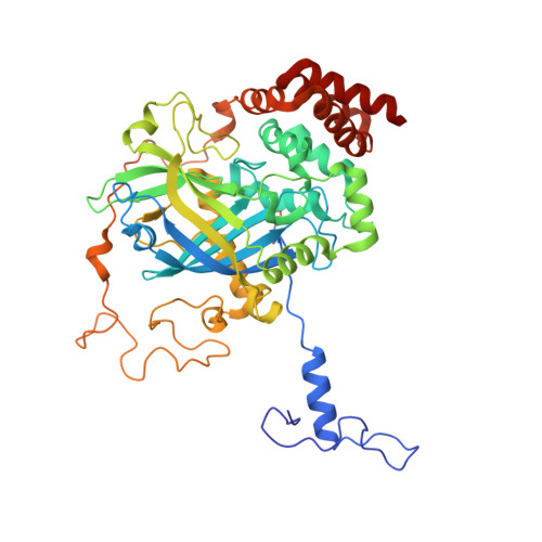
Crystal structure of Proteus mirabilis PR catalase with and without bound NADPH.

Gouet, P.Jouve, H.M.Dideberg, O.

(1995) J Mol Biol 249: 933-954

  • DOI: https://doi.org/10.1006/jmbi.1995.0350
  • Primary Citation of Related Structures:  
    1M85, 2CAH

  • PubMed Abstract: 

    A catalase from a peroxide resistant mutant of Proteus mirabilis binds NADPH tightly. Interestingly, this enzyme can be stripped of NADPH without loss of the catalatic activity. It is the only known non-mammalian catalase able to bind NADPH. The structure without cofactor was solved by molecular replacement using the structure of beef liver catalase as a model. The structure was refined to an R-factor of 19.3% in the range 8 to 2.2 A resolution. According to the sequence, a methionine sulphone was positioned in the haem active site. This oxidized form of methionine is particular to Proteus mirabilis catalase and likely to produce some steric hindrance in the active site. Two important water molecules are positioned in the haem distal site. These two water molecules are not located in the structure of beef liver catalase, but are supposed to account for the catalytic mechanism. The liganded form was obtained by soaking crystals of the unliganded form into an NADPH solution. The structure was refined to an R-factor of 15.9% in the range of 8 to 3.1 A resolution using the unliganded structure as a model. The NADPH was clearly located in the electron density map with the same conformation as in beef liver catalase. The NADPH binding induces slight structural changes. However, the imidazole ring of a histidine residue (His284) rotates about 50 degrees to accommodate the cofactor. The electron transfer from NADPH to the haem molecule was examined and several pathways are proposed.


  • Organizational Affiliation

    Institut de Biologie Structurale Jean-Pierre EBEL 1LCM, Grenoble, France.


Macromolecules
Find similar proteins by:  (by identity cutoff)  |  3D Structure
Entity ID: 1
MoleculeChains Sequence LengthOrganismDetailsImage
catalase484Proteus mirabilisMutation(s): 0 
EC: 1.11.1.6
UniProt
Find proteins for P42321 (Proteus mirabilis)
Explore P42321 
Go to UniProtKB:  P42321
Entity Groups  
Sequence Clusters30% Identity50% Identity70% Identity90% Identity95% Identity100% Identity
UniProt GroupP42321
Sequence Annotations
Expand
  • Reference Sequence
Small Molecules
Ligands 3 Unique
IDChains Name / Formula / InChI Key2D Diagram3D Interactions
HEM
Query on HEM

Download Ideal Coordinates CCD File 
J [auth A]PROTOPORPHYRIN IX CONTAINING FE
C34 H32 Fe N4 O4
KABFMIBPWCXCRK-RGGAHWMASA-L
SO4
Query on SO4

Download Ideal Coordinates CCD File 
B [auth A]
C [auth A]
D [auth A]
E [auth A]
F [auth A]
B [auth A],
C [auth A],
D [auth A],
E [auth A],
F [auth A],
G [auth A],
H [auth A],
I [auth A]
SULFATE ION
O4 S
QAOWNCQODCNURD-UHFFFAOYSA-L
GOL
Query on GOL

Download Ideal Coordinates CCD File 
K [auth A]GLYCEROL
C3 H8 O3
PEDCQBHIVMGVHV-UHFFFAOYSA-N
Modified Residues  1 Unique
IDChains TypeFormula2D DiagramParent
OMT
Query on OMT
A
L-PEPTIDE LINKINGC5 H11 N O4 SMET
Experimental Data & Validation

Experimental Data

  • Method: X-RAY DIFFRACTION
  • Resolution: 2.00 Å
  • R-Value Free: 0.218 
  • R-Value Work: 0.194 
  • R-Value Observed: 0.194 
  • Space Group: P 62 2 2
Unit Cell:
Length ( Å )Angle ( ˚ )
a = 110α = 90
b = 110β = 90
c = 250γ = 120
Software Package:
Software NamePurpose
AMoREphasing
CNSrefinement
MOSFLMdata reduction
CCP4data scaling

Structure Validation

View Full Validation Report


Ligand Structure Quality Assessment 


Entry History 

Deposition Data

Revision History  (Full details and data files)

  • Version 1.0: 2002-08-14
    Type: Initial release
  • Version 1.1: 2008-04-28
    Changes: Version format compliance
  • Version 1.2: 2011-07-13
    Changes: Derived calculations, Version format compliance
  • Version 1.3: 2014-04-16
    Changes: Experimental preparation