Structure and orientation of a G protein fragment in the receptor bound state from residual dipolar couplings.
Koenig, B.W., Kontaxis, G., Mitchell, D.C., Louis, J.M., Litman, B.J., Bax, A.(2002) J Mol Biol 322: 441-461
- PubMed: 12217702
- DOI: https://doi.org/10.1016/s0022-2836(02)00745-3
- Primary Citation of Related Structures:
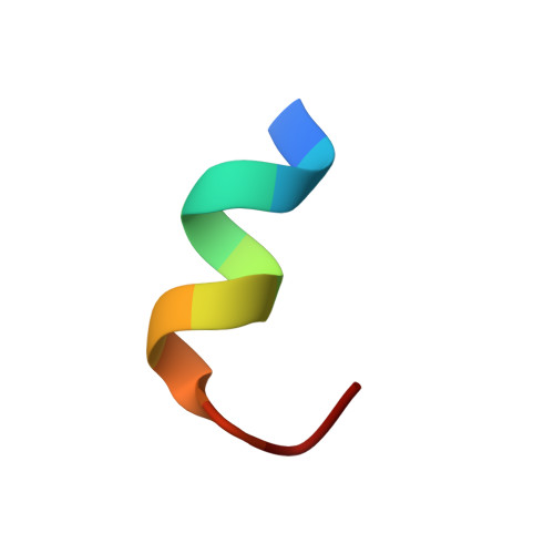
1LVZ - PubMed Abstract:
Residual dipolar couplings for a ligand that is in fast exchange between a free state and a state where it is bound to a macroscopically ordered membrane protein carry precise information on the structure and orientation of the bound ligand. The couplings originate in the bound state but can be detected on the free ligand using standard high resolution NMR. This approach is used to study an analog of the C-terminal undecapeptide of the alpha-subunit of the heterotrimeric G protein transducin when bound to photo-activated rhodopsin. Rhodopsin is the major constituent of disk-shaped membrane vesicles from rod outer segments of bovine retinas, which align spontaneously in the NMR magnet. Photo-activation of rhodopsin triggers transient binding of the peptide, resulting in measurable dipolar contributions to 1J(NH) and 1J(CH) splittings. These dipolar couplings report on the time-averaged orientation of bond vectors in the bound peptide relative to the magnetic field, i.e. relative to the membrane normal. Approximate distance restraints of the bound conformation were derived from transferred NOEs, as measured from the difference of NOESY spectra recorded prior to and after photo-activation. The N-terminal eight residues of the bound undecapeptide adopt a near-ideal alpha-helical conformation. The helix is terminated by an alpha(L) type C-cap, with Gly9 at the C' position in the center of the reverse turn. The angle between the helix axis and the membrane normal is 40 degrees (+/-4) degrees. Peptide protons that make close contact with the receptor are identified by analysis of the NOESY cross-relaxation pattern and include the hydrophobic C terminus of the peptide.
Organizational Affiliation:
Structural Biology Institute, IBI-2, Research Center Jülich, D-52425 Jülich, Germany. b.koenig@fz-juelich.de