Crystal structure of decameric fructose-6-phosphate aldolase from Escherichia coli reveals inter-subunit helix swapping as a structural basis for assembly differences in the transaldolase family.
Thorell, S., Schurmann, M., Sprenger, G.A., Schneider, G.(2002) J Mol Biol 319: 161-171
- PubMed: 12051943
- DOI: https://doi.org/10.1016/S0022-2836(02)00258-9
- Primary Citation of Related Structures:
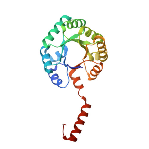
1L6W - PubMed Abstract:
Fructose-6-phosphate aldolase from Escherichia coli is a member of a small enzyme subfamily (MipB/TalC family) that belongs to the class I aldolases. The three-dimensional structure of this enzyme has been determined at 1.93 A resolution by single isomorphous replacement and tenfold non-crystallographic symmetry averaging and refined to an R-factor of 19.9% (R(free) 21.3%). The subunit folds into an alpha/beta barrel, with the catalytic lysine residue on barrel strand beta 4. It is very similar in overall structure to that of bacterial and mammalian transaldolases, although more compact due to extensive deletions of additional secondary structural elements. The enzyme forms a decamer of identical subunits with point group symmetry 52. Five subunits are arranged as a pentamer, and two ring-like pentamers pack like a doughnut to form the decamer. A major interaction within the pentamer is through the C-terminal helix from one monomer, which runs across the active site of the neighbouring subunit. In classical transaldolases, this helix folds back and covers the active site of the same subunit and is involved in dimer formation. The inter-subunit helix swapping appears to be a major determinant for the formation of pentamers rather than dimers while at the same time preserving importing interactions of this helix with the active site of the enzyme. The active site lysine residue is covalently modified, by forming a carbinolamine with glyceraldehyde from the crystallisation mixture. The catalytic machinery is very similar to that of transaldolase, which together with the overall structural similarity suggests that enzymes of the MipB/TALC subfamily are evolutionary related to the transaldolase family.
Organizational Affiliation:
Division of Molecular Structural Biology, Department of Medical Biochemistry and Biophysics, Karolinska Institutet, Tomtebodavägen 6, S-171 77 Stockholm, Sweden.