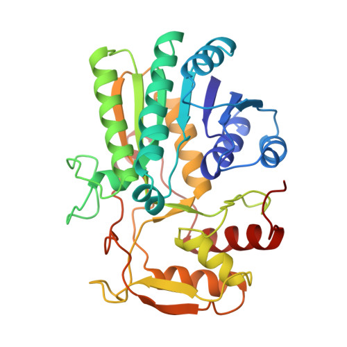
Mechanistic roles of tyrosine 149 and serine 124 in UDP-galactose 4-epimerase from Escherichia coli.
Liu, Y., Thoden, J.B., Kim, J., Berger, E., Gulick, A.M., Ruzicka, F.J., Holden, H.M., Frey, P.A.(1997) Biochemistry 36: 10675-10684
- PubMed: 9271498
- DOI: https://doi.org/10.1021/bi970430a
- Primary Citation of Related Structures:
1KVU - PubMed Abstract:
Synthesis and overexpression of a gene encoding Escherichia coli UDP-galactose 4-epimerase and engineered to facilitate cassette mutagenesis are described. General acid-base catalysis at the active site of this epimerase has been studied by kinetic and spectroscopic analysis of the wild-type enzyme and its specifically mutated forms Y149F, S124A, S124V, and S124T. The X-ray crystal structure of Y149F as its abortive complex with UDP-glucose is structurally similar to that of the corresponding wild-type complex, except for the absence of the phenolic oxygen of Tyr 149. The major effects of mutations are expressed in the values of kcat and kcat/Km. The least active mutant is Y149F, for which the value of kcat is 0.010% of that of the wild-type epimerase. The activity of S124A is also very low, with a kcat value that is 0.035% of that of the native enzyme. The values of Km for Y149F and S124A are 12 and 21% of that of the wild-type enzyme, respectively. The value of kcat for S124T is about 30% of that of the wild-type enzyme, and the value of Km is similar to that of the native enzyme. The reactivities of the mutants in UMP-dependent reductive inactivation by glucose are similarly affected, with kobs being decreased by 6560-, 370-, and 3.4-fold for Y149F, S124A, and S124T, respectively. The second-order rate constants for reductive inactivation by NaBH3CN, which does not require general base catalysis, are similar to that for the native enzyme in the cases of S124A, S124T, and S124V. However, Y149F reacts with NaBH3CN 12-20-fold faster than the wild-type enzyme at pH 8.5 and 7.0, respectively. The increased rate for Y149F is attributed to the weakened charge-transfer interaction between Phe 149 and NAD+, which is present with Tyr 149 in the wild-type enzyme. The charge-transfer band is present in the serine mutants, and its intensity at 320 nm is pH-dependent. The pH dependencies of A320 showed that the pKa values for Tyr 149 are 6.08 for the wild-type epimerase, 6.71 for S124A, 6.86 for S124V, and 6.28 for S124T. The low pKa value for Tyr 149 is attributed mainly to the positive electrostatic field created by NAD+ and Lys 153 (4.5 kcal mol-1) and partly to hydrogen bonding with Ser 124 (1 kcal mol-1). The pKa of Tyr 149 is the same as the kinetic pKa for the Bronsted base that facilitates hydride transfer to NAD+. We concluded that Tyr 149 provides the driving force for general acid-base catalysis, with Ser 124 playing an important role in mediating proton transfer.
Organizational Affiliation:
Institute for Enzyme Research, Graduate School, University of Wisconsin-Madison 53705, USA.