1KEU

The crystal structure of dTDP-D-glucose 4,6-dehydratase (RmlB) from Salmonella enterica serovar Typhimurium with dTDP-D-glucose bound


Experimental Data Snapshot

  • Method: X-RAY DIFFRACTION
  • Resolution: 2.40 Å
  • R-Value Free: 0.222 
  • R-Value Work: 0.197 

wwPDB Validation   3D Report Full Report


This is version 1.4 of the entry. See complete history


Literature

Toward a structural understanding of the dehydratase mechanism.

Allard, S.T.Beis, K.Giraud, M.F.Hegeman, A.D.Gross, J.W.Wilmouth, R.C.Whitfield, C.Graninger, M.Messner, P.Allen, A.G.Maskell, D.J.Naismith, J.H.

(2002) Structure 10: 81-92

  • DOI: https://doi.org/10.1016/s0969-2126(01)00694-3
  • Primary Citation of Related Structures:  
    1KEP, 1KER, 1KET, 1KEU, 1KEW

  • PubMed Abstract: 

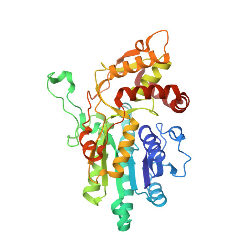
    dTDP-D-glucose 4,6-dehydratase (RmlB) was first identified in the L-rhamnose biosynthetic pathway, where it catalyzes the conversion of dTDP-D-glucose into dTDP-4-keto-6-deoxy-D-glucose. The structures of RmlB from Salmonella enterica serovar Typhimurium in complex with substrate deoxythymidine 5'-diphospho-D-glucose (dTDP-D-glucose) and deoxythymidine 5'-diphosphate (dTDP), and RmlB from Streptococcus suis serotype 2 in complex with dTDP-D-glucose, dTDP, and deoxythymidine 5'-diphospho-D-pyrano-xylose (dTDP-xylose) have all been solved at resolutions between 1.8 A and 2.4 A. The structures show that the active sites are highly conserved. Importantly, the structures show that the active site tyrosine functions directly as the active site base, and an aspartic and glutamic acid pairing accomplishes the dehydration step of the enzyme mechanism. We conclude that the substrate is required to move within the active site to complete the catalytic cycle and that this movement is driven by the elimination of water. The results provide insight into members of the SDR superfamily.


  • Organizational Affiliation

    Centre for Biomolecular Sciences, North Haugh, The University, St. Andrews, Fife KY16 9ST, Scotland, United Kingdom.


Macromolecules
Find similar proteins by:  (by identity cutoff)  |  3D Structure
Entity ID: 1
MoleculeChains Sequence LengthOrganismDetailsImage
dTDP-D-glucose 4,6-dehydratase
A, B
361Salmonella enterica subsp. enterica serovar TyphimuriumMutation(s): 0 
Gene Names: rmlB
EC: 4.2.1.46
UniProt
Find proteins for P26391 (Salmonella typhimurium (strain LT2 / SGSC1412 / ATCC 700720))
Explore P26391 
Go to UniProtKB:  P26391
Entity Groups  
Sequence Clusters30% Identity50% Identity70% Identity90% Identity95% Identity100% Identity
UniProt GroupP26391
Sequence Annotations
Expand
  • Reference Sequence
Small Molecules
Ligands 2 Unique
IDChains Name / Formula / InChI Key2D Diagram3D Interactions
NAD
Query on NAD

Download Ideal Coordinates CCD File 
D [auth A],
F [auth B]
NICOTINAMIDE-ADENINE-DINUCLEOTIDE
C21 H27 N7 O14 P2
BAWFJGJZGIEFAR-NNYOXOHSSA-N
DAU
Query on DAU

Download Ideal Coordinates CCD File 
C [auth A],
E [auth B]
2'DEOXY-THYMIDINE-5'-DIPHOSPHO-ALPHA-D-GLUCOSE
C16 H26 N2 O16 P2
YSYKRGRSMLTJNL-URARBOGNSA-N
Experimental Data & Validation

Experimental Data

  • Method: X-RAY DIFFRACTION
  • Resolution: 2.40 Å
  • R-Value Free: 0.222 
  • R-Value Work: 0.197 
  • Space Group: P 61
Unit Cell:
Length ( Å )Angle ( ˚ )
a = 171.859α = 90
b = 171.859β = 90
c = 94.287γ = 120
Software Package:
Software NamePurpose
AMoREphasing
CNSrefinement
MOSFLMdata reduction
CCP4data scaling

Structure Validation

View Full Validation Report


Entry History 

Deposition Data

Revision History  (Full details and data files)

  • Version 1.0: 2002-01-25
    Type: Initial release
  • Version 1.1: 2008-04-27
    Changes: Version format compliance
  • Version 1.2: 2011-07-13
    Changes: Version format compliance
  • Version 1.3: 2012-05-23
    Changes: Atomic model
  • Version 1.4: 2023-08-16
    Changes: Data collection, Database references, Derived calculations, Refinement description