1KBZ

Crystal Structure of apo-dTDP-6-deoxy-L-lyxo-4-hexulose reductase (RmlD) from Salmonella enterica serovar Typhimurium


Experimental Data Snapshot

  • Method: X-RAY DIFFRACTION
  • Resolution: 2.20 Å
  • R-Value Free: 0.256 
  • R-Value Work: 0.189 
  • R-Value Observed: 0.192 

wwPDB Validation   3D Report Full Report


This is version 1.3 of the entry. See complete history


Literature

Variation on a theme of SDR. dTDP-6-deoxy-L- lyxo-4-hexulose reductase (RmlD) shows a new Mg2+-dependent dimerization mode.

Blankenfeldt, W.Kerr, I.D.Giraud, M.F.McMiken, H.J.Leonard, G.Whitfield, C.Messner, P.Graninger, M.Naismith, J.H.

(2002) Structure 10: 773-786

  • DOI: https://doi.org/10.1016/s0969-2126(02)00770-0
  • Primary Citation of Related Structures:  
    1KBZ, 1KC1, 1KC3, 1N2S

  • PubMed Abstract: 

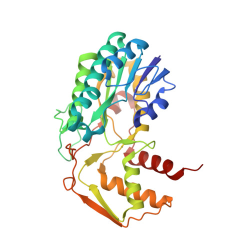
    dTDP-6-deoxy-L-lyxo-4-hexulose reductase (RmlD) catalyzes the final step in the conversion of dTDP-D-glucose to dTDP-L-rhamnose in an NAD(P)H- and Mg2+-dependent reaction. L-rhamnose biosynthesis is an antibacterial target. The structure of RmlD from Salmonella enterica serovar Typhimurium has been determined, and complexes with NADH, NADPH, and dTDP-L-rhamnose are reported. RmlD differs from other short chain dehydrogenases in that it has a novel dimer interface that contains Mg2+. Enzyme catalysis involves hydride transfer from the nicotinamide ring of the cofactor to the C4'-carbonyl group of the substrate. The substrate is activated through protonation by a conserved tyrosine. NAD(P)H is bound in a solvent-exposed cleft, allowing facile replacement. We suggest a novel role for the conserved serine/threonine residue of the catalytic triad of SDR enzymes.


  • Organizational Affiliation

    The Centre for Biomolecular Sciences, The University, St. Andrews, KY16 9ST, Scotland, United Kingdom.


Macromolecules
Find similar proteins by:  (by identity cutoff)  |  3D Structure
Entity ID: 1
MoleculeChains Sequence LengthOrganismDetailsImage
dTDP-glucose oxidoreductase299Salmonella enterica subsp. enterica serovar TyphimuriumMutation(s): 0 
Gene Names: rfbA
EC: 1.1.1.133
UniProt
Find proteins for P26392 (Salmonella typhimurium (strain LT2 / SGSC1412 / ATCC 700720))
Explore P26392 
Go to UniProtKB:  P26392
Entity Groups  
Sequence Clusters30% Identity50% Identity70% Identity90% Identity95% Identity100% Identity
UniProt GroupP26392
Sequence Annotations
Expand
  • Reference Sequence
Small Molecules
Ligands 1 Unique
IDChains Name / Formula / InChI Key2D Diagram3D Interactions
MG
Query on MG

Download Ideal Coordinates CCD File 
B [auth A]MAGNESIUM ION
Mg
JLVVSXFLKOJNIY-UHFFFAOYSA-N
Experimental Data & Validation

Experimental Data

  • Method: X-RAY DIFFRACTION
  • Resolution: 2.20 Å
  • R-Value Free: 0.256 
  • R-Value Work: 0.189 
  • R-Value Observed: 0.192 
  • Space Group: P 21 21 2
Unit Cell:
Length ( Å )Angle ( ˚ )
a = 47.533α = 90
b = 71.901β = 90
c = 82.158γ = 90
Software Package:
Software NamePurpose
SOLVEphasing
REFMACrefinement
MOSFLMdata reduction
CCP4data scaling

Structure Validation

View Full Validation Report


Entry History 

Deposition Data

Revision History  (Full details and data files)

  • Version 1.0: 2002-06-28
    Type: Initial release
  • Version 1.1: 2008-04-27
    Changes: Version format compliance
  • Version 1.2: 2011-07-13
    Changes: Derived calculations, Version format compliance
  • Version 1.3: 2024-02-07
    Changes: Data collection, Database references, Derived calculations