1K8I

CRYSTAL STRUCTURE OF MOUSE H2-DM


Experimental Data Snapshot

  • Method: X-RAY DIFFRACTION
  • Resolution: 3.10 Å
  • R-Value Free: 0.256 
  • R-Value Work: 0.205 
  • R-Value Observed: 0.205 

wwPDB Validation   3D Report Full Report


This is version 1.3 of the entry. See complete history


Literature

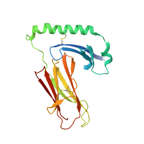
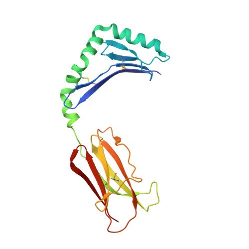
Crystal structure of mouse H2-M.

Fremont, D.H.Crawford, F.Marrack, P.Hendrickson, W.A.Kappler, J.

(1998) Immunity 9: 385-393

  • DOI: https://doi.org/10.1016/s1074-7613(00)80621-4
  • Primary Citation of Related Structures:  
    1K8I

  • PubMed Abstract: 

    H2-M (HLA-DM in humans) resides in an acidic endosomal compartment, where it facilitates the loading of antigenic peptides into the peptide-binding groove of class II MHC. The crystal structure of a soluble form of H2-M has been solved to 3.1 A resolution, revealing a heterodimer with structural similarities to the MHC family of proteins. In contrast to its antigen-presenting cousins, the membrane distal alpha helices of H2-M pack closely together, occluding most of the binding groove except for a single large pocket near the center. The structure of H2-M has several unique features that may play a role in its function as a molecular chaperone and peptide exchange factor.


  • Organizational Affiliation

    Center for Immunology, Department of Pathology, Washington University School of Medicine, St. Louis, Missouri 63110, USA. fremont@immunology.wustl.edu


Macromolecules
Find similar proteins by:  (by identity cutoff)  |  3D Structure
Entity ID: 1
MoleculeChains Sequence LengthOrganismDetailsImage
MHC class II H2-M alpha chain191Mus musculusMutation(s): 0 
Gene Names: H2-DM
UniProt
Find proteins for P28078 (Mus musculus)
Explore P28078 
Go to UniProtKB:  P28078
Entity Groups  
Sequence Clusters30% Identity50% Identity70% Identity90% Identity95% Identity100% Identity
UniProt GroupP28078
Sequence Annotations
Expand
  • Reference Sequence
Find similar proteins by:  (by identity cutoff)  |  3D Structure
Entity ID: 2
MoleculeChains Sequence LengthOrganismDetailsImage
MHC class II H2-M beta 2 chain191Mus musculusMutation(s): 0 
Gene Names: H2-DM
UniProt
Find proteins for Q31099 (Mus musculus)
Explore Q31099 
Go to UniProtKB:  Q31099
Entity Groups  
Sequence Clusters30% Identity50% Identity70% Identity90% Identity95% Identity100% Identity
UniProt GroupQ31099
Sequence Annotations
Expand
  • Reference Sequence
Small Molecules
Ligands 1 Unique
IDChains Name / Formula / InChI Key2D Diagram3D Interactions
NAG
Query on NAG

Download Ideal Coordinates CCD File 
C [auth A],
D [auth A]
2-acetamido-2-deoxy-beta-D-glucopyranose
C8 H15 N O6
OVRNDRQMDRJTHS-FMDGEEDCSA-N
Experimental Data & Validation

Experimental Data

  • Method: X-RAY DIFFRACTION
  • Resolution: 3.10 Å
  • R-Value Free: 0.256 
  • R-Value Work: 0.205 
  • R-Value Observed: 0.205 
  • Space Group: I 4
Unit Cell:
Length ( Å )Angle ( ˚ )
a = 142.7α = 90
b = 142.7β = 90
c = 76.9γ = 90
Software Package:
Software NamePurpose
DENZOdata reduction
SCALEPACKdata scaling
AMoREphasing
X-PLORmodel building
X-PLORrefinement
X-PLORphasing

Structure Validation

View Full Validation Report


Entry History 

Deposition Data

Revision History  (Full details and data files)

  • Version 1.0: 2001-12-05
    Type: Initial release
  • Version 1.1: 2008-04-27
    Changes: Version format compliance
  • Version 1.2: 2011-07-13
    Changes: Non-polymer description, Version format compliance
  • Version 1.3: 2020-07-29
    Type: Remediation
    Reason: Carbohydrate remediation
    Changes: Data collection, Derived calculations, Structure summary