1JHJ

Crystal structure of the APC10/Doc1 subunit of the human anaphase-promoting complex


Experimental Data Snapshot

  • Method: X-RAY DIFFRACTION
  • Resolution: 1.60 Å
  • R-Value Free: 0.244 
  • R-Value Work: 0.209 

wwPDB Validation   3D Report Full Report


This is version 1.5 of the entry. See complete history


Literature

Crystal structure of the APC10/DOC1 subunit of the human anaphase-promoting complex

Wendt, K.S.Vodermaier, H.C.Jacob, U.Gieffers, C.Gmachl, M.Peters, J.M.Huber, R.Sondermann, P.

(2001) Nat Struct Biol 8: 784-788

  • DOI: https://doi.org/10.1038/nsb0901-784
  • Primary Citation of Related Structures:  
    1JHJ

  • PubMed Abstract: 

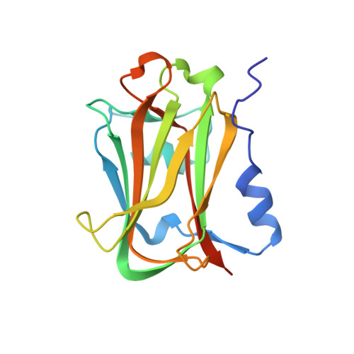
    The anaphase-promoting complex (APC), or cyclosome, is a cell cycle-regulated ubiquitin ligase that controls progression through mitosis and the G1 phase of the cell cycle. The APC is composed of at least 11 subunits; no structure has been determined for any of these subunits. The subunit APC10/DOC1, a one-domain protein consisting of 185 amino acids, has a conserved core (residues 22-161) that is homologous to domains found in several other putative ubiquitin ligases and, therefore, may play a role in ubiquitination reactions. Here we report the crystal structure of human APC10 at 1.6 A resolution. The core of the protein is formed by a beta-sandwich that adopts a jellyroll fold. Unexpectedly, this structure is highly similar to ligand-binding domains of several bacterial and eukaryotic proteins, such as galactose oxidase and coagulation factor Va, raising the possibility that APC10 may function by binding a yet unidentified ligand. We further provide biochemical evidence that the C-terminus of APC10 binds to CDC27/APC3, an APC subunit that contains multiple tetratrico peptide repeats.


  • Organizational Affiliation

    Max-Planck-Institut für Biochemie, Abteilung Strukturforschung, Am Klopferspitz 18a, D-82152 Martinsried, Germany. wendt@biochem.mpg.de


Macromolecules
Find similar proteins by:  (by identity cutoff)  |  3D Structure
Entity ID: 1
MoleculeChains Sequence LengthOrganismDetailsImage
APC10171Homo sapiensMutation(s): 1 
UniProt & NIH Common Fund Data Resources
Find proteins for Q9UM13 (Homo sapiens)
Explore Q9UM13 
Go to UniProtKB:  Q9UM13
PHAROS:  Q9UM13
GTEx:  ENSG00000164162 
Entity Groups  
Sequence Clusters30% Identity50% Identity70% Identity90% Identity95% Identity100% Identity
UniProt GroupQ9UM13
Sequence Annotations
Expand
  • Reference Sequence
Small Molecules
Ligands 1 Unique
IDChains Name / Formula / InChI Key2D Diagram3D Interactions
NI
Query on NI

Download Ideal Coordinates CCD File 
B [auth A]NICKEL (II) ION
Ni
VEQPNABPJHWNSG-UHFFFAOYSA-N
Experimental Data & Validation

Experimental Data

  • Method: X-RAY DIFFRACTION
  • Resolution: 1.60 Å
  • R-Value Free: 0.244 
  • R-Value Work: 0.209 
  • Space Group: P 6 2 2
Unit Cell:
Length ( Å )Angle ( ˚ )
a = 97.664α = 90
b = 97.664β = 90
c = 67.769γ = 120
Software Package:
Software NamePurpose
MLPHAREphasing
CNSrefinement
DENZOdata reduction
SCALEPACKdata scaling

Structure Validation

View Full Validation Report


Entry History 

Deposition Data

Revision History  (Full details and data files)

  • Version 1.0: 2001-10-24
    Type: Initial release
  • Version 1.1: 2008-04-27
    Changes: Version format compliance
  • Version 1.2: 2011-07-13
    Changes: Version format compliance
  • Version 1.3: 2017-03-29
    Changes: Refinement description
  • Version 1.4: 2021-11-10
    Changes: Database references, Derived calculations
  • Version 1.5: 2022-04-13
    Changes: Database references