1J83

STRUCTURE OF FAM17 CARBOHYDRATE BINDING MODULE FROM CLOSTRIDIUM CELLULOVORANS


Experimental Data Snapshot

  • Method: X-RAY DIFFRACTION
  • Resolution: 1.70 Å
  • R-Value Free: 0.219 
  • R-Value Work: 0.198 

wwPDB Validation   3D Report Full Report


This is version 1.2 of the entry. See complete history


Literature

Recognition of cello-oligosaccharides by a family 17 carbohydrate-binding module: an X-ray crystallographic, thermodynamic and mutagenic study.

Notenboom, V.Boraston, A.B.Chiu, P.Freelove, A.C.Kilburn, D.G.Rose, D.R.

(2001) J Mol Biol 314: 797-806

  • DOI: https://doi.org/10.1006/jmbi.2001.5153
  • Primary Citation of Related Structures:  
    1J83, 1J84

  • PubMed Abstract: 

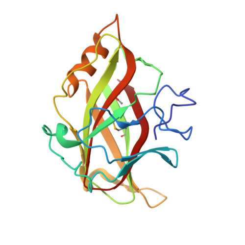
    The crystal structure of the Clostridium cellulovorans carbohydrate-binding module (CBM) belonging to family 17 has been solved to 1.7 A resolution by multiple anomalous dispersion methods. CBM17 binds to non-crystalline cellulose and soluble beta-1,4-glucans, with a minimal binding requirement of cellotriose and optimal affinity for cellohexaose. The crystal structure of CBM17 complexed with cellotetraose solved at 2.0 A resolution revealed that binding occurs in a cleft on the surface of the molecule involving two tryptophan residues and several charged amino acids. Thermodynamic binding studies and alanine scanning mutagenesis in combination with the cellotetraose complex structure allowed the mapping of the CBM17 binding cleft. In contrast to the binding groove characteristic of family 4 CBMs, family 17 CBMs appear to have a very shallow binding cleft that may be more accessible to cellulose chains in non-crystalline cellulose than the deeper binding clefts of family 4 CBMs. The structural differences in these two modules may reflect non-overlapping binding niches on cellulose surfaces.


  • Organizational Affiliation

    Protein Engineering Networks of Centres of Excellence, University of British Columbia, Vancouver, Canada.


Macromolecules
Find similar proteins by:  (by identity cutoff)  |  3D Structure
Entity ID: 1
MoleculeChains Sequence LengthOrganismDetailsImage
ENDO-1,4-BETA GLUCANASE ENGF
A, B
180Clostridium cellulovoransMutation(s): 2 
EC: 3.2.1.4
UniProt
Find proteins for P94622 (Clostridium cellulovorans)
Explore P94622 
Go to UniProtKB:  P94622
Entity Groups  
Sequence Clusters30% Identity50% Identity70% Identity90% Identity95% Identity100% Identity
UniProt GroupP94622
Sequence Annotations
Expand
  • Reference Sequence
Small Molecules
Ligands 1 Unique
IDChains Name / Formula / InChI Key2D Diagram3D Interactions
CA
Query on CA

Download Ideal Coordinates CCD File 
C [auth A],
D [auth B]
CALCIUM ION
Ca
BHPQYMZQTOCNFJ-UHFFFAOYSA-N
Modified Residues  1 Unique
IDChains TypeFormula2D DiagramParent
MSE
Query on MSE
A, B
L-PEPTIDE LINKINGC5 H11 N O2 SeMET
Experimental Data & Validation

Experimental Data

  • Method: X-RAY DIFFRACTION
  • Resolution: 1.70 Å
  • R-Value Free: 0.219 
  • R-Value Work: 0.198 
  • Space Group: C 1 2 1
Unit Cell:
Length ( Å )Angle ( ˚ )
a = 131.264α = 90
b = 37.678β = 91.93
c = 71.234γ = 90
Software Package:
Software NamePurpose
SOLVEphasing
CNSrefinement
DENZOdata reduction
SCALEPACKdata scaling

Structure Validation

View Full Validation Report


Entry History 

Deposition Data

Revision History  (Full details and data files)

  • Version 1.0: 2001-12-12
    Type: Initial release
  • Version 1.1: 2008-04-27
    Changes: Version format compliance
  • Version 1.2: 2011-07-13
    Changes: Version format compliance