1J0N

Crystal Structure of Bacillus sp. GL1 Xanthan Lyase that Acts on Side Chains of Xanthan


Experimental Data Snapshot

  • Method: X-RAY DIFFRACTION
  • Resolution: 2.40 Å
  • R-Value Free: 0.241 
  • R-Value Work: 0.169 
  • R-Value Observed: 0.169 

wwPDB Validation   3D Report Full Report


This is version 1.5 of the entry. See complete history


Literature

Crystal Structure of Bacillus sp. GL1 Xanthan Lyase, Which Acts on the Side Chains of Xanthan.

Hashimoto, W.Nankai, H.Mikami, B.Murata, K.

(2003) J Biol Chem 278: 7663-7673

  • DOI: https://doi.org/10.1074/jbc.M208100200
  • Primary Citation of Related Structures:  
    1J0M, 1J0N

  • PubMed Abstract: 

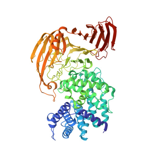
    Xanthan lyase, a member of polysaccharide lyase family 8, is a key enzyme for complete depolymerization of a bacterial heteropolysaccharide, xanthan, in Bacillus sp. GL1. The enzyme acts exolytically on the side chains of the polysaccharide. The x-ray crystallographic structure of xanthan lyase was determined by the multiple isomorphous replacement method. The crystal structures of xanthan lyase and its complex with the product (pyruvylated mannose) were refined at 2.3 and 2.4 A resolution with final R-factors of 17.5 and 16.9%, respectively. The refined structure of the product-free enzyme comprises 752 amino acid residues, 248 water molecules, and one calcium ion. The enzyme consists of N-terminal alpha-helical and C-terminal beta-sheet domains, which constitute incomplete alpha(5)/alpha(5)-barrel and anti-parallel beta-sheet structures, respectively. A deep cleft is located in the N-terminal alpha-helical domain facing the interface between the two domains. Although the overall structure of the enzyme is basically the same as that of the family 8 lyases for hyaluronate and chondroitin AC, significant differences were observed in the loop structure over the cleft. The crystal structure of the xanthan lyase complexed with pyruvylated mannose indicates that the sugar-binding site is located in the deep cleft, where aromatic and positively charged amino acid residues are involved in the binding. The Arg(313) and Tyr(315) residues in the loop from the N-terminal domain and the Arg(612) residue in the loop from the C-terminal domain directly bind to the pyruvate moiety of the product through the formation of hydrogen bonds, thus determining the substrate specificity of the enzyme.


  • Organizational Affiliation

    Department of Basic and Applied Molecular Biotechnology, Division of Food and Biological Science, Graduate School of Agriculture, Kyoto, Uji, Kyoto 611-0011, Japan.


Macromolecules
Find similar proteins by:  (by identity cutoff)  |  3D Structure
Entity ID: 1
MoleculeChains Sequence LengthOrganismDetailsImage
XANTHAN LYASE752Bacillus sp. GL1Mutation(s): 0 
Gene Names: XLY
EC: 4.2.2.12
UniProt
Find proteins for Q9AQS0 (Bacillus sp. (strain GL1))
Explore Q9AQS0 
Go to UniProtKB:  Q9AQS0
Entity Groups  
Sequence Clusters30% Identity50% Identity70% Identity90% Identity95% Identity100% Identity
UniProt GroupQ9AQS0
Sequence Annotations
Expand
  • Reference Sequence
Small Molecules
Ligands 2 Unique
IDChains Name / Formula / InChI Key2D Diagram3D Interactions
CEG
Query on CEG

Download Ideal Coordinates CCD File 
B [auth A]4,6-O-[(1S)-1-carboxyethylidene]-beta-D-glucopyranose
C9 H14 O8
QVVFNJUJKXWFAU-GLPGPYIRSA-N
CA
Query on CA

Download Ideal Coordinates CCD File 
C [auth A]CALCIUM ION
Ca
BHPQYMZQTOCNFJ-UHFFFAOYSA-N
Experimental Data & Validation

Experimental Data

  • Method: X-RAY DIFFRACTION
  • Resolution: 2.40 Å
  • R-Value Free: 0.241 
  • R-Value Work: 0.169 
  • R-Value Observed: 0.169 
  • Space Group: P 21 21 21
Unit Cell:
Length ( Å )Angle ( ˚ )
a = 54.357α = 90
b = 91.305β = 90
c = 159.258γ = 90
Software Package:
Software NamePurpose
FRAMBOdata collection
SAINTdata reduction
PHASESphasing
CNSrefinement
SAINTdata scaling

Structure Validation

View Full Validation Report


Entry History 

Deposition Data

Revision History  (Full details and data files)

  • Version 1.0: 2003-04-01
    Type: Initial release
  • Version 1.1: 2008-04-27
    Changes: Version format compliance
  • Version 1.2: 2011-07-13
    Changes: Source and taxonomy, Version format compliance
  • Version 1.3: 2017-10-04
    Changes: Refinement description
  • Version 1.4: 2020-07-29
    Type: Remediation
    Reason: Carbohydrate remediation
    Changes: Advisory, Database references, Derived calculations, Structure summary
  • Version 1.5: 2023-12-27
    Changes: Data collection, Database references, Structure summary