1ISM

Crystal Structure Analysis of BST-1/CD157 complexed with nicotinamide


Experimental Data Snapshot

  • Method: X-RAY DIFFRACTION
  • Resolution: 3.00 Å
  • R-Value Free: 0.232 
  • R-Value Work: 0.189 

wwPDB Validation   3D Report Full Report


This is version 2.2 of the entry. See complete history


Literature

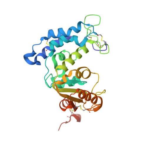
Crystallographic studies on human BST-1/CD157 with ADP-ribosyl cyclase and NAD glycohydrolase activities.

Yamamoto-Katayama, S.Ariyoshi, M.Ishihara, K.Hirano, T.Jingami, H.Morikawa, K.

(2002) J Mol Biol 316: 711-723

  • DOI: https://doi.org/10.1006/jmbi.2001.5386
  • Primary Citation of Related Structures:  
    1ISM

  • PubMed Abstract: 

    cADPR is the novel second messenger that elicits calcium release from intracellular calcium stores and works independently of IP(3). In mammals, the ADP-ribosyl cyclase function is found in two membrane proteins, CD38 and BST-1/CD157. These enzymes, exposed extracellularly, bear cADPR hydrolase and NAD glycohydrolase activities. In spite of its functional importance, the structural basis of these enzymatic reactions remains elusive. We determined the crystal structures of the extracellular region of human BST-1 at atomic resolution in the free form and in complexes with five substrate analogues: nicotinamide, NMN, ATPgammaS, ethenoNADP, and ethenoNAD. The three-dimensional structural views of the reaction centre with these ligands revealed the mode of substrate binding and the catalytic mechanism of the multifunctional enzymatic reactions. In each catalytic cleft of the dimeric enzyme, substrates are recognized predominantly through van der Waals interactions with two tryptophan residues, and thereby the N-glycosidic bond of NAD is correctly exposed near a catalytic glutamate residue. Its carboxyl side-chain stabilizes the catalytic intermediate of the S(N)-1 type reaction. This conformation of the catalytic cleft also implies the mechanism of cyclization between the adenine base and the ribose. The three key residues are invariant among the sequences of BST-1, CD38, and Aplysia cyclase, and hence this substrate recognition mode and catalytic scheme appear to be common in the cyclase family.


  • Organizational Affiliation

    Department of Molecular Biology, Research Institute (BERI), Suita-City, Osaka, Japan.


Macromolecules
Find similar proteins by:  (by identity cutoff)  |  3D Structure
Entity ID: 1
MoleculeChains Sequence LengthOrganismDetailsImage
bone marrow stromal cell antigen 1
A, B
265Homo sapiensMutation(s): 3 
EC: 3.2.2.5
UniProt & NIH Common Fund Data Resources
Find proteins for Q10588 (Homo sapiens)
Explore Q10588 
Go to UniProtKB:  Q10588
PHAROS:  Q10588
GTEx:  ENSG00000109743 
Entity Groups  
Sequence Clusters30% Identity50% Identity70% Identity90% Identity95% Identity100% Identity
UniProt GroupQ10588
Sequence Annotations
Expand
  • Reference Sequence
Oligosaccharides

Help

Entity ID: 2
MoleculeChains Length2D Diagram Glycosylation3D Interactions
2-acetamido-2-deoxy-beta-D-glucopyranose-(1-4)-2-acetamido-2-deoxy-beta-D-glucopyranose
C, D
2N-Glycosylation
Glycosylation Resources
GlyTouCan:  G42666HT
GlyCosmos:  G42666HT
GlyGen:  G42666HT
Experimental Data & Validation

Experimental Data

  • Method: X-RAY DIFFRACTION
  • Resolution: 3.00 Å
  • R-Value Free: 0.232 
  • R-Value Work: 0.189 
  • Space Group: P 21 21 21
Unit Cell:
Length ( Å )Angle ( ˚ )
a = 57.896α = 90
b = 112.728β = 90
c = 133.553γ = 90
Software Package:
Software NamePurpose
DENZOdata reduction
SCALEPACKdata scaling
CNSrefinement
CNSphasing

Structure Validation

View Full Validation Report


Entry History 

Deposition Data

Revision History  (Full details and data files)

  • Version 1.0: 2002-03-13
    Type: Initial release
  • Version 1.1: 2008-04-27
    Changes: Version format compliance
  • Version 1.2: 2011-07-13
    Changes: Non-polymer description, Version format compliance
  • Version 2.0: 2020-07-29
    Type: Remediation
    Reason: Carbohydrate remediation
    Changes: Atomic model, Data collection, Derived calculations, Structure summary
  • Version 2.1: 2021-11-10
    Changes: Database references, Structure summary
  • Version 2.2: 2023-12-27
    Changes: Data collection