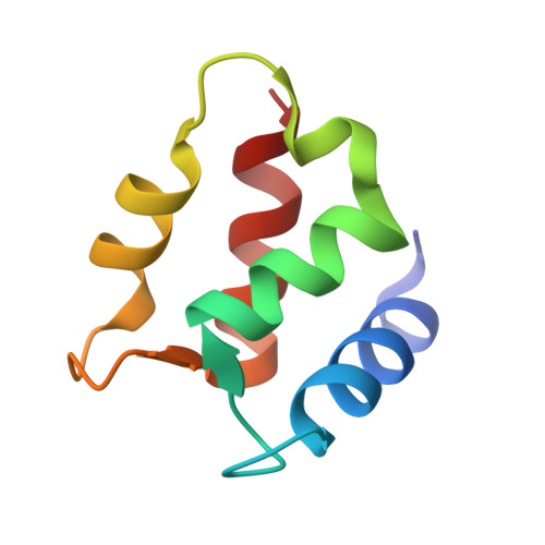
1IGV

BOVINE CALBINDIN D9K BINDING MN2+


Experimental Data Snapshot

  • Method: X-RAY DIFFRACTION
  • Resolution: 1.85 Å
  • R-Value Free: 0.272 
  • R-Value Work: 0.186 

wwPDB Validation   3D Report Full Report


This is version 1.4 of the entry. See complete history


Literature

Structural basis for the negative allostery between Ca(2+)- and Mg(2+)-binding in the intracellular Ca(2+)-receptor calbindin D9k.

Andersson, M.Malmendal, A.Linse, S.Ivarsson, I.Forsen, S.Svensson, L.A.

(1997) Protein Sci 6: 1139-1147

  • DOI: https://doi.org/10.1002/pro.5560060602
  • Primary Citation of Related Structures:  
    1IG5, 1IGV

  • PubMed Abstract: 

    The three-dimensional structures of the magnesium- and manganese-bound forms of calbindin D9k were determined to 1.6 A and 1.9 A resolution, respectively, using X-ray crystallography. These two structures are nearly identical but deviate significantly from both the calcium bound form and the metal ion-free (apo) form. The largest structural differences are seen in the C-terminal EF-hand, and involve changes in both metal ion coordination and helix packing. The N-terminal calcium binding site is not occupied by any metal ion in the magnesium and manganese structures, and shows little structural deviation from the apo and calcium bound forms. 1H-NMR and UV spectroscopic studies at physiological ion concentrations show that the C-terminal site of the protein is significantly populated by magnesium at resting cell calcium levels, and that there is a negative allosteric interaction between magnesium and calcium binding. Calcium binding was found to occur with positive cooperativity at physiological magnesium concentration.


  • Organizational Affiliation

    Department of Molecular Biophysics, Lund University, Sweden.


Macromolecules
Find similar proteins by:  (by identity cutoff)  |  3D Structure
Entity ID: 1
MoleculeChains Sequence LengthOrganismDetailsImage
VITAMIN D-DEPENDENT CALCIUM-BINDING PROTEIN, INTESTINAL75Bos taurusMutation(s): 0 
Gene Names: synthetic gene
UniProt
Find proteins for P02633 (Bos taurus)
Explore P02633 
Go to UniProtKB:  P02633
Entity Groups  
Sequence Clusters30% Identity50% Identity70% Identity90% Identity95% Identity100% Identity
UniProt GroupP02633
Sequence Annotations
Expand
  • Reference Sequence
Small Molecules
Ligands 1 Unique
IDChains Name / Formula / InChI Key2D Diagram3D Interactions
MN
Query on MN

Download Ideal Coordinates CCD File 
B [auth A]MANGANESE (II) ION
Mn
WAEMQWOKJMHJLA-UHFFFAOYSA-N
Experimental Data & Validation

Experimental Data

  • Method: X-RAY DIFFRACTION
  • Resolution: 1.85 Å
  • R-Value Free: 0.272 
  • R-Value Work: 0.186 
  • Space Group: P 43 21 2
Unit Cell:
Length ( Å )Angle ( ˚ )
a = 33.8α = 90
b = 33.8β = 90
c = 129.63γ = 90
Software Package:
Software NamePurpose
SIGMAAmodel building
X-PLORrefinement
XDSdata reduction
AUTOMARdata reduction
SIGMAAphasing

Structure Validation

View Full Validation Report


Entry History 

Deposition Data

Revision History  (Full details and data files)

  • Version 1.0: 2001-04-25
    Type: Initial release
  • Version 1.1: 2008-04-27
    Changes: Version format compliance
  • Version 1.2: 2011-07-13
    Changes: Version format compliance
  • Version 1.3: 2017-10-04
    Changes: Refinement description
  • Version 1.4: 2024-02-07
    Changes: Data collection, Database references, Derived calculations