Molecular models and structural comparisons of native and mutant class I filamentous bacteriophages Ff (fd, f1, M13), If1 and IKe.
Marvin, D.A., Hale, R.D., Nave, C., Helmer-Citterich, M.(1994) J Mol Biol 235: 260-286
- PubMed: 8289247
- DOI: https://doi.org/10.1016/s0022-2836(05)80032-4
- Primary Citation of Related Structures:
1IFI, 1IFJ, 1IFK, 1IFL - PubMed Abstract:
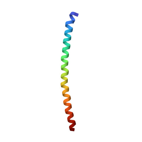
The filamentous bacteriophages are flexible rods about 1 to 2 microns long and 6 nm in diameter, with a helical shell of protein subunits surrounding a DNA core. The approximately 50-residue coat protein subunit is largely alpha-helix and the axis of the alpha-helix makes a small angle with the axis of the virion. The protein shell can be considered in three sections: the outer surface, occupied by the N-terminal region of the subunit, rich in acidic residues that interact with the surrounding solvent and give the virion a low isoelectric point; the interior of the shell, including a 19-residue stretch of apolar side-chains, where protein subunits interact mainly with each other; and the inner surface, occupied by the C-terminal region of the subunit, rich in basic residues that interact with the DNA core. The fact that virtually all protein side-chain interactions are between different subunits in the coat protein array, rather than within subunits, makes this a useful model system for studies of interactions between alpha-helix subunits in a macromolecular assembly. We describe molecular models of the class I filamentous bacteriophages. This class includes strains fd, f1, M13 (these 3 very similar strains are members of the Ff group), If1 and IKe. Our model of fd has been refined to fit quantitative X-ray fibre diffraction data to 30 A resolution in the meridional direction and 7 A resolution in the equatorial direction. A simulated 3.3 A resolution diffraction pattern from this model has the same general distribution of intensity as the experimental diffraction pattern. The observed diffraction data at 7 A resolution are fitted much better by the calculated diffraction pattern of our molecular model than by that of a model in which the alpha-helix subunit is represented by a rod of uniform density. The fact that our fd model explains the fd diffraction data is only part of our structure analysis. The atomic details of the model are supported by non-diffraction data, in part previously published and in part newly reported here. These data include information about permitted or forbidden side-chain replacements, about the effect of chemical modification, and about spectroscopic experiments.(ABSTRACT TRUNCATED AT 400 WORDS)
Organizational Affiliation:
Department of Biochemistry, University of Cambridge, U.K.