1IAL

IMPORTIN ALPHA, MOUSE


Experimental Data Snapshot

  • Method: X-RAY DIFFRACTION
  • Resolution: 2.50 Å
  • R-Value Free: 0.240 
  • R-Value Work: 0.189 
  • R-Value Observed: 0.189 

wwPDB Validation   3D Report Full Report


This is version 1.3 of the entry. See complete history


Literature

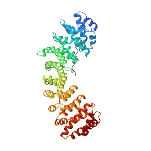
Autoinhibition by an internal nuclear localization signal revealed by the crystal structure of mammalian importin alpha.

Kobe, B.

(1999) Nat Struct Biol 6: 388-397

  • DOI: https://doi.org/10.1038/7625
  • Primary Citation of Related Structures:  
    1IAL

  • PubMed Abstract: 

    Importin alpha is the nuclear import receptor that recognizes classical monopartite and bipartite nuclear localization signals (NLSs). The structure of mouse importin alpha has been determined at 2.5 A resolution. The structure shows a large C-terminal domain containing armadillo repeats, and a less structured N-terminal importin beta-binding domain containing an internal NLS bound to the NLS-binding site. The structure explains the regulatory switch between the cytoplasmic, high-affinity form, and the nuclear, low-affinity form for NLS binding of the nuclear import receptor predicted by the current models of nuclear import. Importin beta conceivably converts the low- to high-affinity form by binding to a site overlapping the autoinhibitory sequence. The structure also has implications for understanding NLS recognition, and the structures of armadillo and HEAT repeats.


  • Organizational Affiliation

    Structural Biology Laboratory, St. Vincent's Institute of Medical Research, Fitzroy, Victoria, Australia. B.Kobe@medicine.unimelb.edu.au


Macromolecules
Find similar proteins by:  (by identity cutoff)  |  3D Structure
Entity ID: 1
MoleculeChains Sequence LengthOrganismDetailsImage
IMPORTIN ALPHA453Mus musculusMutation(s): 0 
UniProt & NIH Common Fund Data Resources
Find proteins for P52293 (Mus musculus)
Explore P52293 
Go to UniProtKB:  P52293
IMPC:  MGI:103561
Entity Groups  
Sequence Clusters30% Identity50% Identity70% Identity90% Identity95% Identity100% Identity
UniProt GroupP52293
Sequence Annotations
Expand
  • Reference Sequence
Experimental Data & Validation

Experimental Data

  • Method: X-RAY DIFFRACTION
  • Resolution: 2.50 Å
  • R-Value Free: 0.240 
  • R-Value Work: 0.189 
  • R-Value Observed: 0.189 
  • Space Group: P 21 21 21
Unit Cell:
Length ( Å )Angle ( ˚ )
a = 78.549α = 90
b = 89.675β = 90
c = 100.562γ = 90
Software Package:
Software NamePurpose
X-PLORmodel building
X-PLORrefinement
DENZOdata reduction
SCALEPACKdata scaling
X-PLORphasing

Structure Validation

View Full Validation Report


Entry History 

Deposition Data

  • Released Date: 1999-06-15 
  • Deposition Author(s): Kobe, B.

Revision History  (Full details and data files)

  • Version 1.0: 1999-06-15
    Type: Initial release
  • Version 1.1: 2008-03-24
    Changes: Version format compliance
  • Version 1.2: 2011-07-13
    Changes: Version format compliance
  • Version 1.3: 2023-08-09
    Changes: Database references, Other, Refinement description