Dynamic characterization of the water binding loop in the P-type cardiotoxin: implication for the role of the bound water molecule.
Sue, S.C., Jarrell, H.C., Brisson, J.R., Wu, W.G.(2001) Biochemistry 40: 12782-12794
- PubMed: 11669614
- DOI: https://doi.org/10.1021/bi010848f
- Primary Citation of Related Structures:
1I02 - PubMed Abstract:
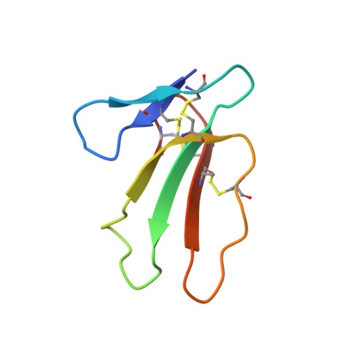
Recent studies of cobra P-type cardiotoxins (CTXs) have shown that the water-binding loop (loop II) plays a crucial role in toxin binding to biological membranes and in their cytotoxicity. To understand the role of bound water in the loop, the structure and dynamics of the major P-type CTX from Taiwan cobra, CTX A3, were determined by a comprehensive NMR analysis involving (1)H NOESY/ROESY, (13)C[1)H]NOE/T(1) relaxation, and (17)O triple-quantum filtered NMR. A single water molecule was found to be tightly hydrogen bonded to the NH of Met26 with a correlation time (5-7 ns) approaching the isotropic tumbling time (3.8-4.5 ns) of the CTX A3 molecule. Surprisingly, despite the relatively long residence time (ca. 5 ns to 100 micros), the bound water molecule of CTX A3 is located within a dynamic (order parameter S(2) approximately 0.7) and solvent accessible loop. Comparison among several P-type CTXs suggests that proline residues in the consensus sequence of MxAxPxVPV should play an important role in the formation of the water binding loop. It is proposed that the exchange rate of the bound water may play a role in regulating the lipid binding mode of amphiphilic CTX molecules near membrane surfaces.
Organizational Affiliation:
Department of Life Sciences, National Tsing Hua University, Hsinchu 30043, Taiwan.