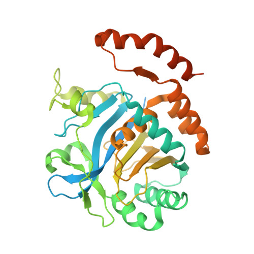
1HYN

CRYSTAL STRUCTURE OF THE CYTOPLASMIC DOMAIN OF HUMAN ERYTHROCYTE BAND-3 PROTEIN


Experimental Data Snapshot

  • Method: X-RAY DIFFRACTION
  • Resolution: 2.60 Å
  • R-Value Free: 0.290 
  • R-Value Work: 0.216 

wwPDB Validation   3D Report Full Report


This is version 1.4 of the entry. See complete history


Literature

Crystallographic structure and functional interpretation of the cytoplasmic domain of erythrocyte membrane band 3.

Zhang, D.Kiyatkin, A.Bolin, J.T.Low, P.S.

(2000) Blood 96: 2925-2933

  • Primary Citation of Related Structures:  
    1HYN

  • PubMed Abstract: 

    The red blood cell membrane (RBCM) is a primary model for animal cell plasma membranes. One of its major organizing centers is the cytoplasmic domain of band 3 (cdb3), which links multiple proteins to the membrane. Included among its peripheral protein ligands are ankyrin (the major bridge to the spectrin-actin skeleton), protein 4. 1, protein 4.2, aldolase, glyceraldehyde-3-phosphate dehydrogenase, phosphofructokinase, deoxyhemoglobin, p72syk protein tyrosine kinase, and hemichromes. The crystal structure of cdb3 is reported at 0.26 nm (2.6 A) resolution. A tight symmetric dimer is formed by cdb3; it is stabilized by interlocked dimerization arms contributed by both monomers. Each subunit also includes a larger peripheral protein binding domain with an alpha(+) beta-fold. The binding sites of several peripheral proteins are localized in the structure, and the nature of the major conformational change that regulates membrane-skeletal interactions is evaluated. An improved structural definition of the protein network at the inner surface of the RBCM is now possible.


  • Organizational Affiliation

    Departments of Chemistry and Biological Sciences, Purdue University, West Lafayette, IN 47907-1392, USA.


Macromolecules
Find similar proteins by:  (by identity cutoff)  |  3D Structure
Entity ID: 1
MoleculeChains Sequence LengthOrganismDetailsImage
BAND 3 ANION TRANSPORT PROTEINA [auth P],
B [auth Q],
C [auth R],
D [auth S]
379Homo sapiensMutation(s): 0 
Membrane Entity: Yes 
UniProt & NIH Common Fund Data Resources
Find proteins for P02730 (Homo sapiens)
Explore P02730 
Go to UniProtKB:  P02730
PHAROS:  P02730
GTEx:  ENSG00000004939 
Entity Groups  
Sequence Clusters30% Identity50% Identity70% Identity90% Identity95% Identity100% Identity
UniProt GroupP02730
Sequence Annotations
Expand
  • Reference Sequence
Experimental Data & Validation

Experimental Data

  • Method: X-RAY DIFFRACTION
  • Resolution: 2.60 Å
  • R-Value Free: 0.290 
  • R-Value Work: 0.216 
  • Space Group: C 1 2 1
Unit Cell:
Length ( Å )Angle ( ˚ )
a = 181.59α = 90
b = 92.58β = 131.86
c = 123.33γ = 90
Software Package:
Software NamePurpose
SOLVEphasing
X-PLORrefinement
DENZOdata reduction
SCALEPACKdata scaling

Structure Validation

View Full Validation Report


Entry History 

Deposition Data

Revision History  (Full details and data files)

  • Version 1.0: 2001-05-16
    Type: Initial release
  • Version 1.1: 2008-04-27
    Changes: Version format compliance
  • Version 1.2: 2011-07-13
    Changes: Version format compliance
  • Version 1.3: 2017-10-04
    Changes: Refinement description
  • Version 1.4: 2024-02-07
    Changes: Data collection, Database references