Crystal Structure of Sedl and its Implications for a Genetic Disease Spondyloepiphyseal Dysplasia Tarda
Jang, S.B., Kim, Y.-G., Cho, Y.-S., Suh, P.-G., Kim, K.-H., Oh, B.-H.(2002) J Biol Chem 277: 49863
- PubMed: 12361953
- DOI: https://doi.org/10.1074/jbc.M207436200
- Primary Citation of Related Structures:
1H3Q - PubMed Abstract:
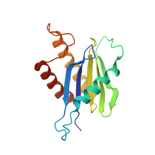
SEDL is an evolutionarily highly conserved protein in eukaryotic organisms. Deletions or point mutations in the SEDL gene are responsible for the genetic disease spondyloepiphyseal dysplasia tarda (SEDT), an X-linked skeletal disorder. SEDL has been identified as a component of the transport protein particle (TRAPP), critically involved in endoplasmic reticulum-to-Golgi vesicle transport. Herein, we report the 2.4 A resolution structure of SEDL, which reveals an unexpected similarity to the structures of the N-terminal regulatory domain of two SNAREs, Ykt6p and Sec22b, despite no sequence homology to these proteins. The similarity and the presence of unusually many solvent-exposed apolar residues of SEDL suggest that it serves regulatory and/or adaptor functions through multiple protein-protein interactions. Of the four known missense mutations responsible for SEDT, three mutations (S73L, F83S, V130D) map to the protein interior, where the mutations would disrupt the structure, and the fourth (D47Y) on a surface at which the mutation may abrogate functional interactions with a partner protein.
Organizational Affiliation:
National Creative Research Initiative Center for Biomolecular Recognition and the Department of Life Science, Pohang University of Science and Technology, Pohang, Kyungbuk 790-784, Korea.