1H36

Structures of Human Oxidosqualene Cyclase Inhibitors Bound to an Homologous Enzyme


Experimental Data Snapshot

  • Method: X-RAY DIFFRACTION
  • Resolution: 2.80 Å
  • R-Value Free: 0.258 
  • R-Value Work: 0.221 
  • R-Value Observed: 0.221 

wwPDB Validation   3D Report Full Report


Ligand Structure Quality Assessment 


This is version 1.3 of the entry. See complete history


Literature

Binding Structures and Potencies of Oxidosqualene Cyclase Inhibitors with the Homologous Squalene-Hopene Cyclase

Lenhart, A.Reinert, D.J.Aebi, J.D.Dehmlow, H.Morand, O.H.Schulz, G.E.

(2003) J Med Chem 46: 2083

  • DOI: https://doi.org/10.1021/jm0211218
  • Primary Citation of Related Structures:  
    1H35, 1H36, 1H37, 1H39, 1H3A, 1H3B, 1H3C, 1O6H, 1O6Q, 1O6R, 1O79

  • PubMed Abstract: 

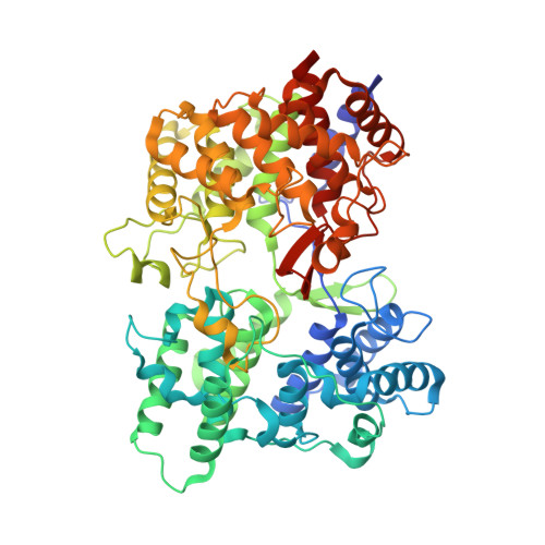
    The binding structures of 11 human oxidosqualene cyclase inhibitors designed as cholesterol-lowering agents were determined for the squalene-hopene cyclase from Alicyclobacillus acidocaldarius, which is the only structurally known homologue of the human enzyme. The complexes were produced by cocrystallization, and the structures were elucidated by X-ray diffraction analyses. All inhibitors were bound in the large active center cavity. The detailed binding structures are presented and discussed in the light of the IC50 values of these 11 as well as 17 other inhibitors. They provide a consistent picture for the inhibition of the bacterial enzyme and can be used to adjust and improve homology models of the human enzyme. The detailed active center structures of the two enzymes are too different to show an IC50 correlation.


  • Organizational Affiliation

    Institut für Organische Chemie und Biochemie, Albert-Ludwigs-Universität, Albertstrasse 21, Freiburg im Breisgau 79104, Germany.


Macromolecules
Find similar proteins by:  (by identity cutoff)  |  3D Structure
Entity ID: 1
MoleculeChains Sequence LengthOrganismDetailsImage
SQUALENE--HOPENE CYCLASE
A, B, C
631Alicyclobacillus acidocaldariusMutation(s): 0 
EC: 5.4.99.17
UniProt
Find proteins for P33247 (Alicyclobacillus acidocaldarius subsp. acidocaldarius (strain ATCC 27009 / DSM 446 / BCRC 14685 / JCM 5260 / KCTC 1825 / NBRC 15652 / NCIMB 11725 / NRRL B-14509 / 104-IA))
Explore P33247 
Go to UniProtKB:  P33247
Entity Groups  
Sequence Clusters30% Identity50% Identity70% Identity90% Identity95% Identity100% Identity
UniProt GroupP33247
Sequence Annotations
Expand
  • Reference Sequence
Small Molecules
Binding Affinity Annotations 
IDSourceBinding Affinity
R88 BindingDB:  1H36 IC50: 18 (nM) from 1 assay(s)
PDBBind:  1H36 IC50: 18 (nM) from 1 assay(s)
Experimental Data & Validation

Experimental Data

  • Method: X-RAY DIFFRACTION
  • Resolution: 2.80 Å
  • R-Value Free: 0.258 
  • R-Value Work: 0.221 
  • R-Value Observed: 0.221 
  • Space Group: P 32 2 1
Unit Cell:
Length ( Å )Angle ( ˚ )
a = 141.513α = 90
b = 141.513β = 90
c = 244.908γ = 120
Software Package:
Software NamePurpose
CNSrefinement
XDSdata reduction
SCALAdata scaling

Structure Validation

View Full Validation Report


Ligand Structure Quality Assessment 


Entry History 

Deposition Data

Revision History  (Full details and data files)

  • Version 1.0: 2003-08-21
    Type: Initial release
  • Version 1.1: 2013-11-20
    Changes: Derived calculations, Non-polymer description, Other, Refinement description, Source and taxonomy, Structure summary, Version format compliance
  • Version 1.2: 2014-02-05
    Changes: Source and taxonomy
  • Version 1.3: 2018-06-13
    Changes: Data collection, Structure summary