A Novel Iron Centre in the Split-Soret Cytochrome C from Desulfovibrio Desulfuricans Atcc 27774
Abreu, I.A., Lourenco, A.I., Xavier, A.V., Legall, J., Coelho, A.V., Matias, P.M., Pinto, D.M., Armenia Carrondo, M., Teixeira, M., Saraiva, L.M.(2003) J Biol Inorg Chem 8: 360
- PubMed: 12589573
- DOI: https://doi.org/10.1007/s00775-002-0426-3
- Primary Citation of Related Structures:
1H21 - PubMed Abstract:
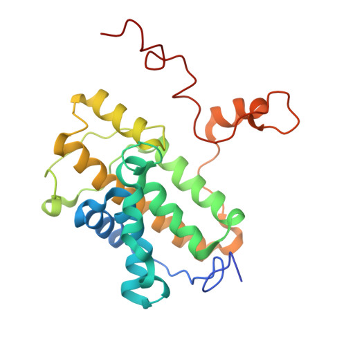
The facultative sulfate/nitrate-reducing bacterium Desulfovibrio desulfuricans ATCC 27774 harbours a split-Soret cytochrome c. This cytochrome is a homodimeric protein, having two bis-histidinyl c-type haems per monomer. It has an unique architecture at the haem domain: each haem has one of the coordinating histidines provided by the other monomer, and in each monomer the haems are parallel to each other, almost in van der Waals contact. This work reports the cloning and sequencing of the gene encoding for this cytochrome and shows, by transcriptional analysis, that it is more expressed in nitrate-grown cells than in sulfate-grown ones. In addition, the gene-deduced amino acid sequence revealed two new cysteine residues that could be involved in the binding of a non-haem iron centre. Indeed, the presence of a novel type of an iron-sulfur centre (possibly of the [2Fe-2S] type) was demonstrated by EPR spectroscopy, and putative models for its localization and structure in the cytochrome molecule are proposed on the basis of the so-far-known 3D crystallographic structure of the aerobically purified split-Soret cytochrome, which lacks this centre.
Organizational Affiliation:
Instituto de Tecnologia Química e Biológica, Universidade Nova de Lisboa, Rua da Quinta Grande 6, 2780-156, Oeiras, Portugal.