Protein crystallization by rational mutagenesis of surface residues: Lys to Ala mutations promote crystallization of RhoGDI.
Longenecker, K.L., Garrard, S.M., Sheffield, P.J., Derewenda, Z.S.(2001) Acta Crystallogr D Biol Crystallogr 57: 679-688
- PubMed: 11320308
- DOI: https://doi.org/10.1107/s0907444901003122
- Primary Citation of Related Structures:
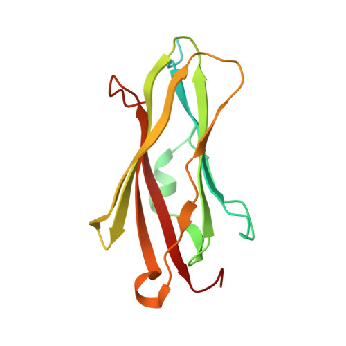
1FSO, 1FST, 1FT0, 1FT3 - PubMed Abstract:
Crystallization is a unique process that occurs at the expense of entropy, including the conformational entropy of surface residues, which become ordered in crystal lattices during formation of crystal contacts. It could therefore be argued that epitopes free of amino acids with high conformational entropy are more thermodynamically favorable for crystal formation. For a protein recalcitrant to crystallization, mutation of such surface amino acids to residues with no conformational entropy might lead to enhancement of crystallization. This paper reports the results of experiments with an important cytosolic regulator of GTPases, human RhoGDI, in which lysine residues were systematically mutated to alanines. Single and multiple mutations were introduced into two different variants of RhoGDI, NDelta23 and NDelta66, in which the first 23 and 66 residues, respectively, were removed by recombinant methods. In total, 13 single and multiple mutants were prepared and assessed for crystallization and all were shown to crystallize using the Hampton Research Crystal Screens I and II, in contrast to wild-type NDelta23 and NDelta66 RhoGDI which did not crystallize. Four crystal structures were solved (the triple mutants NDelta23:K135,138,141A and NDelta66:K135,138,141A, and two single mutants NDelta66:K113A and NDelta66:K141A) and in three cases the crystal contacts of the new lattices were found precisely at the sites of mutations. These results support the notion that it is, in principle, possible to rationally design mutations which systematically enhance proteins' ability to crystallize.
Organizational Affiliation:
Department of Molecular Physiology and Biological Physics, University of Virginia, PO Box 800736, Charlottesville, VA 22908-0736, USA.