1ETE

CRYSTAL STRUCTURE OF THE FLT3 LIGAND


Experimental Data Snapshot

  • Method: X-RAY DIFFRACTION
  • Resolution: 2.20 Å
  • R-Value Free: 0.287 
  • R-Value Work: 0.239 

wwPDB Validation   3D Report Full Report


This is version 1.2 of the entry. See complete history


Literature

Flt3 ligand structure and unexpected commonalities of helical bundles and cystine knots.

Savvides, S.N.Boone, T.Andrew Karplus, P.

(2000) Nat Struct Biol 7: 486-491

  • DOI: https://doi.org/10.1038/75896
  • Primary Citation of Related Structures:  
    1ETE

  • PubMed Abstract: 

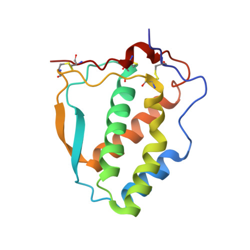
    Human Flt3 ligand (Flt3L) stimulates early hematopoiesis by activating a type III tyrosine kinase receptor on primitive bone marrow stem cells. The crystal structure of soluble Flt3L reveals that it is a homodimer of two short chain alpha-helical bundles. Comparisons of structure-function relationships of Flt3L with the homologous hematopoietic cytokines macrophage colony stimulating factor (MCSF) and stem cell factor (SCF) suggest that they have a common receptor binding mode that is distinct from the paradigm derived from the complex of growth hormone with its receptor. Furthermore, we identify recognition features common to all helical and cystine-knot protein ligands that activate type III tyrosine kinase receptors, and the closely related type V tyrosine kinase receptors.


  • Organizational Affiliation

    Program in Biophysics, Cornell University, Ithaca, NY 14853, USA.


Macromolecules
Find similar proteins by:  (by identity cutoff)  |  3D Structure
Entity ID: 1
MoleculeChains Sequence LengthOrganismDetailsImage
FLT3 LIGAND
A, B, C, D
134Homo sapiensMutation(s): 0 
UniProt & NIH Common Fund Data Resources
Find proteins for P49771 (Homo sapiens)
Explore P49771 
Go to UniProtKB:  P49771
GTEx:  ENSG00000090554 
Entity Groups  
Sequence Clusters30% Identity50% Identity70% Identity90% Identity95% Identity100% Identity
UniProt GroupP49771
Sequence Annotations
Expand
  • Reference Sequence
Experimental Data & Validation

Experimental Data

  • Method: X-RAY DIFFRACTION
  • Resolution: 2.20 Å
  • R-Value Free: 0.287 
  • R-Value Work: 0.239 
  • Space Group: P 21 21 2
Unit Cell:
Length ( Å )Angle ( ˚ )
a = 137.08α = 90
b = 159.41β = 90
c = 26.25γ = 90
Software Package:
Software NamePurpose
DENZOdata reduction
SCALEPACKdata scaling
PHASESphasing
X-PLORrefinement

Structure Validation

View Full Validation Report


Entry History 

Deposition Data

Revision History  (Full details and data files)

  • Version 1.0: 2000-06-14
    Type: Initial release
  • Version 1.1: 2008-04-27
    Changes: Version format compliance
  • Version 1.2: 2011-07-13
    Changes: Version format compliance