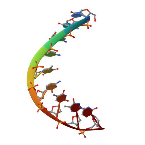
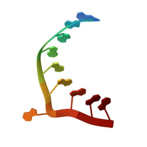
1EFO

CRYSTAL STRUCTURE OF AN ADENINE BULGE IN THE RNA CHAIN OF A DNA/RNA HYBRID, D(CTCCTCTTC)/R(GAAGAGAGAG)


Experimental Data Snapshot

  • Method: X-RAY DIFFRACTION
  • Resolution: 2.30 Å
  • R-Value Free: 0.222 
  • R-Value Work: 0.199 
  • R-Value Observed: 0.199 

wwPDB Validation   3D Report Full Report


This is version 1.4 of the entry. See complete history


Literature

Crystal structure of an adenine bulge in the RNA chain of a DNA.RNA hybrid, d(CTCCTCTTC).r(gaagagagag).

Sudarsanakumar, C.Xiong, Y.Sundaralingam, M.

(2000) J Mol Biol 299: 103-112

  • DOI: https://doi.org/10.1006/jmbi.2000.3730
  • Primary Citation of Related Structures:  
    1EFO

  • PubMed Abstract: 

    Crystal structure of a DNA.RNA hybrid, d(CTCCTCTTC).r(gaagagagag), with an adenine bulge in the polypurine RNA strand was determined at 2.3 A resolution. The structure was solved by the molecular replacement method and refined to a final R-factor of 19.9% (Rfree 22.2%). The hybrid duplex crystallized in the space group I222 with unit cell dimensions, a = 46.66 A, b = 47.61 A and c = 54.05 A, and adopts the A-form conformation. All RNA and DNA sugars are in the C3'-endo conformation, the glycosyl angles in anti conformation and the majority of the C4'-C5' torsion angles in g+ except two trans angles, in conformity with the C3'-endo rigid nucleotide hypothesis. The adenine bulge is looped out and it is also in the anti C3'-endo conformation. The bulge is involved in a base-triple (C.g)*a interaction with the end base-pair (C9.g10) in the minor groove of a symmetry-related molecule. The 2' hydroxyl group of g15 is hydrogen bonded to O2P and O5' of g17, skipping the bulged adenine a16 and stabilizing the sugar-phosphate backbone of the hybrid. The hydrogen bonding and the backbone conformation at the bulged adenine site is very similar to that found in the crystal structure of a protein-RNA complex.


  • Organizational Affiliation

    Department of Chemistry, Biochemistry, Ohio State University, Columbus 43210, USA.


Macromolecules

Find similar nucleic acids by:  Sequence   |   3D Structure  

Entity ID: 1
MoleculeChains LengthOrganismImage
DNA (5'-D(*CP*TP*CP*CP*TP*CP*TP*TP*C)-3')9N/A
Sequence Annotations
Expand
  • Reference Sequence

Find similar nucleic acids by:  Sequence   |   3D Structure  

Entity ID: 2
MoleculeChains LengthOrganismImage
RNA (5'-R(*GP*AP*AP*GP*AP*GP*AP*GP*AP*G)-3')10N/A
Sequence Annotations
Expand
  • Reference Sequence
Experimental Data & Validation

Experimental Data

  • Method: X-RAY DIFFRACTION
  • Resolution: 2.30 Å
  • R-Value Free: 0.222 
  • R-Value Work: 0.199 
  • R-Value Observed: 0.199 
  • Space Group: I 2 2 2
Unit Cell:
Length ( Å )Angle ( ˚ )
a = 46.66α = 90
b = 47.61β = 90
c = 54.04γ = 90
Software Package:
Software NamePurpose
AMoREphasing
CNSrefinement
SCALEPACKdata scaling

Structure Validation

View Full Validation Report


Entry History 

Deposition Data

Revision History  (Full details and data files)

  • Version 1.0: 2000-05-29
    Type: Initial release
  • Version 1.1: 2008-04-27
    Changes: Version format compliance
  • Version 1.2: 2011-07-13
    Changes: Version format compliance
  • Version 1.3: 2017-10-04
    Changes: Refinement description
  • Version 1.4: 2024-02-07
    Changes: Data collection, Database references