1EEH

UDP-N-ACETYLMURAMOYL-L-ALANINE:D-GLUTAMATE LIGASE


Experimental Data Snapshot

  • Method: X-RAY DIFFRACTION
  • Resolution: 1.90 Å
  • R-Value Free: 0.265 
  • R-Value Work: 0.219 
  • R-Value Observed: 0.226 

wwPDB Validation   3D Report Full Report


Ligand Structure Quality Assessment 


This is version 1.5 of the entry. See complete history


Literature

"Open" structures of MurD: domain movements and structural similarities with folylpolyglutamate synthetase.

Bertrand, J.A.Fanchon, E.Martin, L.Chantalat, L.Auger, G.Blanot, D.van Heijenoort, J.Dideberg, O.

(2000) J Mol Biol 301: 1257-1266

  • DOI: https://doi.org/10.1006/jmbi.2000.3994
  • Primary Citation of Related Structures:  
    1E0D, 1EEH

  • PubMed Abstract: 

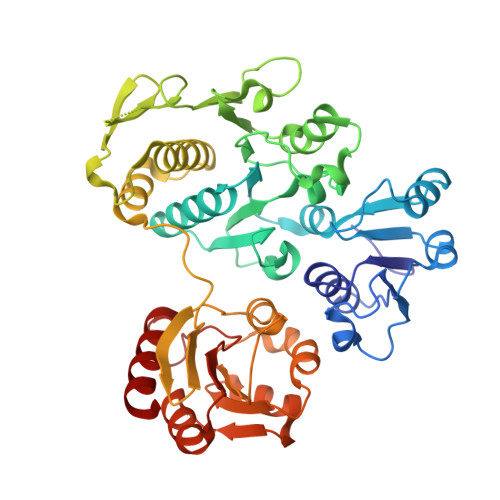
    UDP-N-acetylmuramoyl-l-alanine:d-glutamate (MurD) ligase catalyses the addition of d-glutamate to the nucleotide precursor UDP-N-acetylmuramoyl-l-alanine (UMA). The crystal structures of Escherichia coli in the substrate-free form and MurD complexed with UMA have been determined at 2.4 A and 1.88 A resolution, respectively. The MurD structure comprises three domains each of a topology reminiscent of nucleotide-binding folds. In the two structures the C-terminal domain undergoes a large rigid-body rotation away from the N-terminal and central domains. These two "open" structures were compared with the four published "closed" structures of MurD. In addition the comparison reveals which regions are affected by the binding of UMA, ATP and d-Glu. Also we compare and discuss two structurally characterized enzymes which belong to the same ligase superfamily: MurD and folylpolyglutamate synthetase (FGS). The analysis allows the identification of key residues involved in the reaction mechanism of FGS. The determination of the two "open" conformation structures represents a new step towards the complete elucidation of the enzymatic mechanism of the MurD ligase.


  • Organizational Affiliation

    Laboratoire de Cristallographie Macromoléculaire, Institut de Biologie Structurale Jean-Pierre Ebel (CNRS-CEA), 41, rue Jules Horowitz, Grenoble, Cedex 1, F-38027, France.


Macromolecules
Find similar proteins by:  (by identity cutoff)  |  3D Structure
Entity ID: 1
MoleculeChains Sequence LengthOrganismDetailsImage
UDP-N-ACETYLMURAMOYL-L-ALANINE:D-GLUTAMATE LIGASE437Escherichia coliMutation(s): 0 
EC: 6.3.2.9
UniProt
Find proteins for P14900 (Escherichia coli (strain K12))
Explore P14900 
Go to UniProtKB:  P14900
Entity Groups  
Sequence Clusters30% Identity50% Identity70% Identity90% Identity95% Identity100% Identity
UniProt GroupP14900
Sequence Annotations
Expand
  • Reference Sequence
Small Molecules
Ligands 1 Unique
IDChains Name / Formula / InChI Key2D Diagram3D Interactions
UMA
Query on UMA

Download Ideal Coordinates CCD File 
B [auth A]URIDINE-5'-DIPHOSPHATE-N-ACETYLMURAMOYL-L-ALANINE
C23 H36 N4 O20 P2
NTMMCWJNQNKACG-KBKUWGQMSA-N
Experimental Data & Validation

Experimental Data

  • Method: X-RAY DIFFRACTION
  • Resolution: 1.90 Å
  • R-Value Free: 0.265 
  • R-Value Work: 0.219 
  • R-Value Observed: 0.226 
  • Space Group: P 21 21 21
Unit Cell:
Length ( Å )Angle ( ˚ )
a = 58.84α = 90
b = 63.28β = 90
c = 114.91γ = 90
Software Package:
Software NamePurpose
AMoREphasing
CNSrefinement
XDSdata reduction
XDSdata scaling

Structure Validation

View Full Validation Report


Ligand Structure Quality Assessment 


Entry History 

Deposition Data

Revision History  (Full details and data files)

  • Version 1.0: 2001-01-17
    Type: Initial release
  • Version 1.1: 2008-04-27
    Changes: Version format compliance
  • Version 1.2: 2011-07-13
    Changes: Version format compliance
  • Version 1.3: 2018-04-04
    Changes: Data collection
  • Version 1.4: 2018-04-11
    Changes: Data collection
  • Version 1.5: 2024-02-07
    Changes: Data collection, Database references, Derived calculations