Inaugural article: retrostructural analysis of metalloproteins: application to the design of a minimal model for diiron proteins.
Lombardi, A., Summa, C.M., Geremia, S., Randaccio, L., Pavone, V., DeGrado, W.F.(2000) Proc Natl Acad Sci U S A 97: 6298-6305
- PubMed: 10841536
- DOI: https://doi.org/10.1073/pnas.97.12.6298
- Primary Citation of Related Structures:
1EC5 - PubMed Abstract:
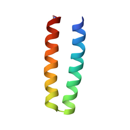
De novo protein design provides an attractive approach for the construction of models to probe the features required for function of complex metalloproteins. The metal-binding sites of many metalloproteins lie between multiple elements of secondary structure, inviting a retrostructural approach to constructing minimal models of their active sites. The backbone geometries comprising the metal-binding sites of zinc fingers, diiron proteins, and rubredoxins may be described to within approximately 1 A rms deviation by using a simple geometric model with only six adjustable parameters. These geometric models provide excellent starting points for the design of metalloproteins, as illustrated in the construction of Due Ferro 1 (DF1), a minimal model for the Glu-Xxx-Xxx-His class of dinuclear metalloproteins. This protein was synthesized and structurally characterized as the di-Zn(II) complex by x-ray crystallography, by using data that extend to 2.5 A. This four-helix bundle protein is comprised of two noncovalently associated helix-loop-helix motifs. The dinuclear center is formed by two bridging Glu and two chelating Glu side chains, as well as two monodentate His ligands. The primary ligands are mostly buried in the protein interior, and their geometries are stabilized by a network of hydrogen bonds to second-shell ligands. In particular, a Tyr residue forms a hydrogen bond to a chelating Glu ligand, similar to a motif found in the diiron-containing R2 subunit of Escherichia coli ribonucleotide reductase and the ferritins. DF1 also binds cobalt and iron ions and should provide an attractive model for a variety of diiron proteins that use oxygen for processes including iron storage, radical formation, and hydrocarbon oxidation.
Organizational Affiliation:
Department of Chemistry, University of Napoli "Federico II," Via Mezzocannone, 4, I-80134 Napoli, Italy.