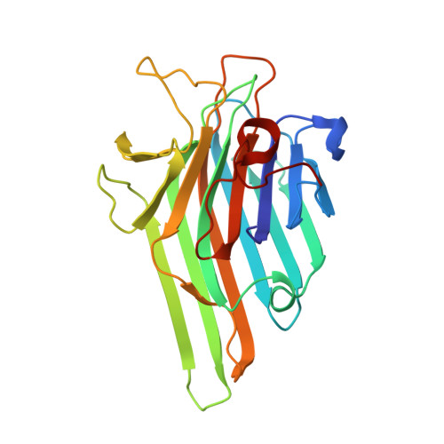
The structural features of concanavalin A governing non-proline peptide isomerization
Bouckaert, J., Dewallef, Y., Poortmans, F., Wyns, L., Loris, R.(2000) J Biol Chem 275: 19778-19787
- PubMed: 10748006
- DOI: https://doi.org/10.1074/jbc.M001251200
- Primary Citation of Related Structures:
1DQ0, 1DQ1, 1DQ2, 1DQ4, 1DQ5, 1DQ6 - PubMed Abstract:
The reversible binding of manganese and calcium to concanavalin A determines the carbohydrate binding of the lectin by inducing large conformational changes. These changes are governed by the isomerization of a non-proline peptide bond, Ala-207-Asp-208, positioned in a beta-strand in between the calcium binding site S2 and the carbohydrate specificity-determining loop. The replacement of calcium by manganese allowed us to investigate the structures of the carbohydrate binding, locked state and the inactive, unlocked state of concanavalin A, both with and without metal ions bound. Crystals of unlocked metal-free concanavalin A convert to the locked form with the binding of two Mn(2+) ions. Removal of these ions from the crystals traps metal-free concanavalin A in its locked state, a minority species in solution. The ligation of a metal ion in S2 to unlocked concanavalin A causes bending of the beta-strand foregoing the S2 ligand residues Asp-10 and Tyr-12. This bending disrupts conventional beta-sheet hydrogen bonding and forces the Thr-11 side chain against the Ala-207-Asp-208 peptide bond. The steric strain exerted by Thr-11 is presumed to drive the trans-to-cis isomerization. Upon isomerization, Asp-208 flips into its carbohydrate binding position, and the conformation of the carbohydrate specificity determining loop changes dramatically.
Organizational Affiliation:
Laboratorium voor Ultrastructuur, Vlaams Interuniversitair Instituut voor Biotechnologie, Vrije Universiteit Brussel, Paardenstraat 65, B-1640 Sint-Genesius-Rode, Belgium. bouckaej@vub.ac.be