1D5Y

CRYSTAL STRUCTURE OF THE E. COLI ROB TRANSCRIPTION FACTOR IN COMPLEX WITH DNA


Experimental Data Snapshot

  • Method: X-RAY DIFFRACTION
  • Resolution: 2.70 Å
  • R-Value Free: 0.302 
  • R-Value Work: 0.254 
  • R-Value Observed: 0.254 

wwPDB Validation   3D Report Full Report


This is version 1.3 of the entry. See complete history


Literature

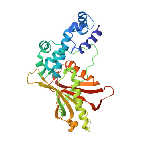
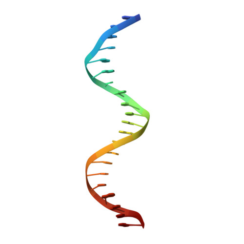
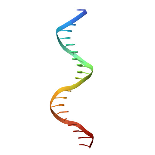
Crystal structure of the Escherichia coli Rob transcription factor in complex with DNA.

Kwon, H.J.Bennik, M.H.Demple, B.Ellenberger, T.

(2000) Nat Struct Biol 7: 424-430

  • DOI: https://doi.org/10.1038/75213
  • Primary Citation of Related Structures:  
    1D5Y

  • PubMed Abstract: 

    The Escherichia coli Rob protein is a transcription factor belonging to the AraC/XylS protein family that regulates genes involved in resistance to antibiotics, organic solvents and heavy metals. The genes encoding these proteins are activated by the homologous proteins MarA and SoxS, although the level of activation can vary for the different transcription factors. Here we report a 2.7 A crystal structure of Rob in complex with the micF promoter that reveals an unusual mode of binding to DNA. The Rob-DNA complex differs from the previously reported structure of MarA bound to the mar promoter, in that only one of Rob's dual helix-turn-helix (HTH) motifs engages the major groove of the binding site. Biochemical studies show that sequence specific interactions involving only one of Rob's HTH motifs are sufficient for high affinity binding to DNA. The two different modes of DNA binding seen in crystal structures of Rob and MarA also match the distinctive patterns of DNA protection by AraC at several sites within the pBAD promoter. These and other findings suggest that gene activation by AraC/XylS transcription factors might involve two alternative modes of binding to DNA in different promoter contexts.


  • Organizational Affiliation

    Graduate Program in Biophysics, Harvard University, Cambridge, MA 02138, USA.


Macromolecules

Find similar proteins by:  (by identity cutoff)  |  3D Structure
Entity ID: 3
MoleculeChains Sequence LengthOrganismDetailsImage
ROB TRANSCRIPTION FACTORE [auth A],
F [auth D],
G [auth C],
H [auth B]
292Escherichia coliMutation(s): 0 
UniProt
Find proteins for P0ACI0 (Escherichia coli (strain K12))
Explore P0ACI0 
Go to UniProtKB:  P0ACI0
Entity Groups  
Sequence Clusters30% Identity50% Identity70% Identity90% Identity95% Identity100% Identity
UniProt GroupP0ACI0
Sequence Annotations
Expand
  • Reference Sequence

Find similar nucleic acids by:  Sequence   |   3D Structure  

Entity ID: 1
MoleculeChains LengthOrganismImage
DNA (5'-D(*TP*GP*AP*CP*AP*GP*CP*AP*CP*TP*GP*AP*AP*TP*GP*TP*CP*AP*AP*AP*G)-3')A [auth M],
C [auth O]
21N/A
Sequence Annotations
Expand
  • Reference Sequence

Find similar nucleic acids by:  Sequence   |   3D Structure  

Entity ID: 2
MoleculeChains LengthOrganismImage
DNA (5'-D(*AP*CP*TP*TP*TP*GP*AP*CP*AP*TP*TP*CP*AP*GP*TP*GP*CP*TP*GP*TP*C)-3')B [auth N],
D [auth P]
21N/A
Sequence Annotations
Expand
  • Reference Sequence
Experimental Data & Validation

Experimental Data

  • Method: X-RAY DIFFRACTION
  • Resolution: 2.70 Å
  • R-Value Free: 0.302 
  • R-Value Work: 0.254 
  • R-Value Observed: 0.254 
  • Space Group: P 1 21 1
Unit Cell:
Length ( Å )Angle ( ˚ )
a = 75.384α = 90
b = 208.012β = 90
c = 67.731γ = 90
Software Package:
Software NamePurpose
MLPHAREphasing
CNSrefinement
DENZOdata reduction
SCALEPACKdata scaling

Structure Validation

View Full Validation Report


Entry History 

Deposition Data

Revision History  (Full details and data files)

  • Version 1.0: 2000-04-24
    Type: Initial release
  • Version 1.1: 2008-04-27
    Changes: Version format compliance
  • Version 1.2: 2011-07-13
    Changes: Version format compliance
  • Version 1.3: 2024-02-07
    Changes: Data collection, Database references