1CM0

CRYSTAL STRUCTURE OF THE PCAF/COENZYME-A COMPLEX


Experimental Data Snapshot

  • Method: X-RAY DIFFRACTION
  • Resolution: 2.30 Å
  • R-Value Free: 0.268 
  • R-Value Work: 0.223 

wwPDB Validation   3D Report Full Report


This is version 1.4 of the entry. See complete history


Literature

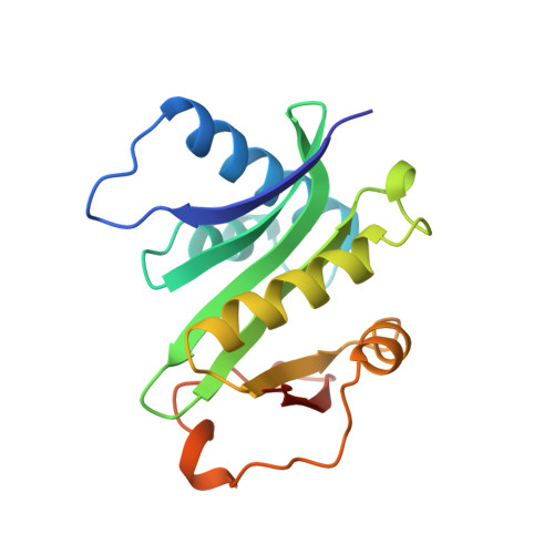
Crystal structure of the histone acetyltransferase domain of the human PCAF transcriptional regulator bound to coenzyme A.

Clements, A.Rojas, J.R.Trievel, R.C.Wang, L.Berger, S.L.Marmorstein, R.

(1999) EMBO J 18: 3521-3532

  • DOI: https://doi.org/10.1093/emboj/18.13.3521
  • Primary Citation of Related Structures:  
    1CM0

  • PubMed Abstract: 

    The human p300/CBP-associating factor, PCAF, mediates transcriptional activation through its ability to acetylate nucleosomal histone substrates as well as transcriptional activators such as p53. We have determined the 2.3 A crystal structure of the histone acetyltransferase (HAT) domain of PCAF bound to coenzyme A. The structure reveals a central protein core associated with coenzyme A binding and a pronounced cleft that sits over the protein core and is flanked on opposite sides by the N- and C-terminal protein segments. A correlation of the structure with the extensive mutagenesis data for PCAF and the homologous yeast GCN5 protein implicates the cleft and the N- and C-terminal protein segments as playing an important role in histone substrate binding, and a glutamate residue in the protein core as playing an essential catalytic role. A structural comparison with the coenzyme-bound forms of the related N-acetyltransferases, HAT1 (yeast histone acetyltransferase 1) and SmAAT (Serratia marcescens aminoglycoside 3-N-acetyltransferase), suggests the mode of substrate binding and catalysis by these enzymes and establishes a paradigm for understanding the structure-function relationships of other enzymes that acetylate histones and transcriptional regulators to promote activated transcription.


  • Organizational Affiliation

    The Wistar Institute, University of Pennsylvania, Philadelphia, PA 19104, USA.


Macromolecules
Find similar proteins by:  (by identity cutoff)  |  3D Structure
Entity ID: 1
MoleculeChains Sequence LengthOrganismDetailsImage
P300/CBP ASSOCIATING FACTORA [auth B],
B [auth A]
168Homo sapiensMutation(s): 0 
UniProt & NIH Common Fund Data Resources
Find proteins for Q92831 (Homo sapiens)
Explore Q92831 
Go to UniProtKB:  Q92831
PHAROS:  Q92831
GTEx:  ENSG00000114166 
Entity Groups  
Sequence Clusters30% Identity50% Identity70% Identity90% Identity95% Identity100% Identity
UniProt GroupQ92831
Sequence Annotations
Expand
  • Reference Sequence
Small Molecules
Ligands 1 Unique
IDChains Name / Formula / InChI Key2D Diagram3D Interactions
COA
Query on COA

Download Ideal Coordinates CCD File 
C [auth B],
D [auth A]
COENZYME A
C21 H36 N7 O16 P3 S
RGJOEKWQDUBAIZ-IBOSZNHHSA-N
Binding Affinity Annotations 
IDSourceBinding Affinity
COA BindingDB:  1CM0 IC50: 4.19e+4 (nM) from 1 assay(s)
Experimental Data & Validation

Experimental Data

  • Method: X-RAY DIFFRACTION
  • Resolution: 2.30 Å
  • R-Value Free: 0.268 
  • R-Value Work: 0.223 
  • Space Group: P 64
Unit Cell:
Length ( Å )Angle ( ˚ )
a = 97α = 90
b = 97β = 90
c = 77.85γ = 120
Software Package:
Software NamePurpose
AMoREphasing
CNSrefinement
DENZOdata reduction
SCALEPACKdata scaling

Structure Validation

View Full Validation Report


Entry History 

Deposition Data

Revision History  (Full details and data files)

  • Version 1.0: 1999-07-06
    Type: Initial release
  • Version 1.1: 2008-04-26
    Changes: Version format compliance
  • Version 1.2: 2011-07-13
    Changes: Version format compliance
  • Version 1.3: 2023-12-27
    Changes: Data collection, Database references, Derived calculations
  • Version 1.4: 2024-04-03
    Changes: Refinement description