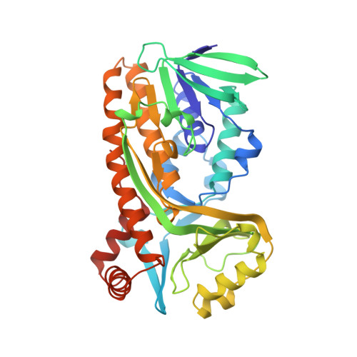
1CC6

PHE161 AND ARG166 VARIANTS OF P-HYDROXYBENZOATE HYDROXYLASE. IMPLICATIONS FOR NADPH RECOGNITION AND STRUCTURAL STABILITY.


Experimental Data Snapshot

  • Method: X-RAY DIFFRACTION
  • Resolution: 2.20 Å
  • R-Value Work: 0.185 

wwPDB Validation   3D Report Full Report


Ligand Structure Quality Assessment 


This is version 1.6 of the entry. See complete history


Literature

Phe161 and Arg166 variants of p-hydroxybenzoate hydroxylase. Implications for NADPH recognition and structural stability.

Eppink, M.H.Bunthol, C.Schreuder, H.A.van Berkel, W.J.

(1999) FEBS Lett 443: 251-255

  • DOI: https://doi.org/10.1016/s0014-5793(98)01726-8
  • Primary Citation of Related Structures:  
    1CC4, 1CC6

  • PubMed Abstract: 

    Phe161 and Arg166 of p-hydroxybenzoate hydroxylase from Pseudomonas fluorescens belong to a newly discovered sequence motif in flavoprotein hydroxylases with a putative dual function in FAD and NADPH binding [1]. To study their role in more detail, Phe161 and Arg166 were selectively changed by site-directed mutagenesis. F161A and F161G are catalytically competent enzymes having a rather poor affinity for NADPH. The catalytic properties of R166K are similar to those of the native enzyme. R166S and R166E show impaired NADPH binding and R166E has lost the ability to bind FAD. The crystal structure of substrate complexed F161A at 2.2 A is indistinguishable from the native enzyme, except for small changes at the site of mutation. The crystal structure of substrate complexed R166S at 2.0 A revealed that Arg166 is important for providing an intimate contact between the FAD binding domain and a long excursion of the substrate binding domain. It is proposed that this interaction is essential for structural stability and for the recognition of the pyrophosphate moiety of NADPH.


  • Organizational Affiliation

    Department of Biomolecular Sciences, Wageningen University Research Centre, The Netherlands.


Macromolecules
Find similar proteins by:  (by identity cutoff)  |  3D Structure
Entity ID: 1
MoleculeChains Sequence LengthOrganismDetailsImage
PROTEIN (P-HYDROXYBENZOATE HYDROXYLASE)394Pseudomonas fluorescensMutation(s): 2 
Gene Names: POBA
EC: 1.14.13.2
UniProt
Find proteins for P00438 (Pseudomonas fluorescens)
Explore P00438 
Go to UniProtKB:  P00438
Entity Groups  
Sequence Clusters30% Identity50% Identity70% Identity90% Identity95% Identity100% Identity
UniProt GroupP00438
Sequence Annotations
Expand
  • Reference Sequence
Small Molecules
Ligands 2 Unique
IDChains Name / Formula / InChI Key2D Diagram3D Interactions
FAD
Query on FAD

Download Ideal Coordinates CCD File 
B [auth A]FLAVIN-ADENINE DINUCLEOTIDE
C27 H33 N9 O15 P2
VWWQXMAJTJZDQX-UYBVJOGSSA-N
PHB
Query on PHB

Download Ideal Coordinates CCD File 
C [auth A]P-HYDROXYBENZOIC ACID
C7 H6 O3
FJKROLUGYXJWQN-UHFFFAOYSA-N
Binding Affinity Annotations 
IDSourceBinding Affinity
PHB Binding MOAD:  1CC6 Kd: 3.00e+4 (nM) from 1 assay(s)
Experimental Data & Validation

Experimental Data

  • Method: X-RAY DIFFRACTION
  • Resolution: 2.20 Å
  • R-Value Work: 0.185 
  • Space Group: C 2 2 21
Unit Cell:
Length ( Å )Angle ( ˚ )
a = 72.2α = 90
b = 146.8β = 90
c = 88.9γ = 90
Software Package:
Software NamePurpose
X-PLORmodel building
X-PLORrefinement
XDSdata reduction
XSCALEdata scaling
X-PLORphasing

Structure Validation

View Full Validation Report


Ligand Structure Quality Assessment 


Entry History 

Deposition Data

Revision History  (Full details and data files)

  • Version 1.0: 1999-03-12
    Type: Initial release
  • Version 1.1: 2007-10-16
    Changes: Version format compliance
  • Version 1.2: 2011-07-13
    Changes: Version format compliance
  • Version 1.3: 2011-12-07
    Changes: Database references
  • Version 1.4: 2017-11-29
    Changes: Derived calculations
  • Version 1.5: 2019-11-06
    Changes: Data collection, Database references
  • Version 1.6: 2023-08-09
    Changes: Data collection, Database references, Derived calculations, Refinement description