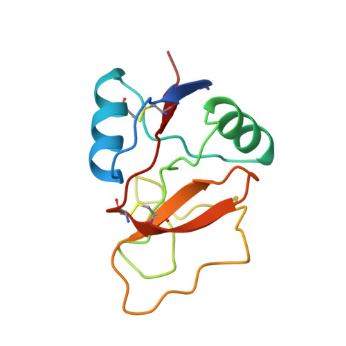
Ca2+-dependent structural changes in C-type mannose-binding proteins.
Ng, K.K., Park-Snyder, S., Weis, W.I.(1998) Biochemistry 37: 17965-17976
- PubMed: 9922165
- DOI: https://doi.org/10.1021/bi981972a
- Primary Citation of Related Structures:
1BUU, 1BV4 - PubMed Abstract:
C-type animal lectins are a diverse family of proteins which mediate cell-surface carbohydrate-recognition events through a conserved carbohydrate-recognition domain (CRD). Most members of this family possess a carbohydrate-binding activity that depends strictly on the binding of Ca2+ at two sites, designated 1 and 2, in the CRD. The structural transitions associated with Ca2+ binding in C-type lectins have been investigated by determining high-resolution crystal structures of rat serum mannose-binding protein (MBP) bound to one Ho3+ in place of Ca2+, and the apo form of rat liver MBP. The removal of Ca2+ does not affect the core structure of the CRD, but dramatic conformational changes occur in the loops. The most significant structural change in the absence of Ca2+ is the isomerization of a cis-peptide bond preceding a conserved proline residue in Ca2+ site 2. This bond adopts the cis conformation in all Ca2+-bound structures, whereas both cis and trans conformations are observed in the absence of Ca2+. The pattern of structural changes in the three loops that interact with Ca2+ is dictated in large part by the conformation of the prolyl peptide bond. The highly conserved nature of Ca2+ site 2 suggests that the transitions observed in MBPs are general features of Ca2+ binding in C-type lectins.
Organizational Affiliation:
Department of Structural Biology, Stanford University School of Medicine, California 94305, USA.