1B12

CRYSTAL STRUCTURE OF TYPE 1 SIGNAL PEPTIDASE FROM ESCHERICHIA COLI IN COMPLEX WITH A BETA-LACTAM INHIBITOR


Experimental Data Snapshot

  • Method: X-RAY DIFFRACTION
  • Resolution: 1.95 Å
  • R-Value Free: 0.246 
  • R-Value Work: 0.220 
  • R-Value Observed: 0.220 

wwPDB Validation   3D Report Full Report


This is version 1.4 of the entry. See complete history


Literature

Crystal structure of a bacterial signal peptidase in complex with a beta-lactam inhibitor.

Paetzel, M.Dalbey, R.E.Strynadka, N.C.

(1998) Nature 396: 186-190

  • DOI: https://doi.org/10.1038/24196
  • Primary Citation of Related Structures:  
    1B12

  • PubMed Abstract: 

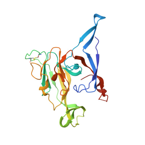
    The signal peptidase (SPase) from Escherichia coli is a membrane-bound endopeptidase with two amino-terminal transmembrane segments and a carboxy-terminal catalytic region which resides in the periplasmic space. SPase functions to release proteins that have been translocated into the inner membrane from the cell interior, by cleaving off their signal peptides. We report here the X-ray crystal structure of a catalytically active soluble fragment of E. coli SPase (SPase delta2-75). We have determined this structure at 1.9 A resolution in a complex with an inhibitor, a beta-lactam (5S,6S penem), which is covalently bound as an acyl-enzyme intermediate to the gamma-oxygen of a serine residue at position 90, demonstrating that this residue acts as the nucleophile in the hydrolytic mechanism of signal-peptide cleavage. The structure is consistent with the use by SPase of Lys 145 as a general base in the activation of the nucleophilic Ser90, explains the specificity requirement at the signal-peptide cleavage site, and reveals a large exposed hydrophobic surface which could be a site for an intimate association with the membrane. As enzymes that are essential for cell viability, bacterial SPases present a feasible antibacterial target: our determination of the SPase structure therefore provides a template for the rational design of antibiotic compounds.


  • Organizational Affiliation

    Department of Biochemistry and Molecular Biology, University of British Columbia, Vancouver, Canada.


Macromolecules
Find similar proteins by:  (by identity cutoff)  |  3D Structure
Entity ID: 1
MoleculeChains Sequence LengthOrganismDetailsImage
SIGNAL PEPTIDASE I
A, B, C, D
248Escherichia coli BL21(DE3)Mutation(s): 0 
Gene Names: LEPB
EC: 3.4.21.89
Membrane Entity: Yes 
UniProt
Find proteins for P00803 (Escherichia coli (strain K12))
Explore P00803 
Go to UniProtKB:  P00803
Entity Groups  
Sequence Clusters30% Identity50% Identity70% Identity90% Identity95% Identity100% Identity
UniProt GroupP00803
Sequence Annotations
Expand
  • Reference Sequence
Small Molecules
Ligands 2 Unique
IDChains Name / Formula / InChI Key2D Diagram3D Interactions
1PN
Query on 1PN

Download Ideal Coordinates CCD File 
E [auth A],
G [auth B],
H [auth C],
I [auth D]
prop-2-en-1-yl (2S)-2-[(2S,3R)-3-(acetyloxy)-1-oxobutan-2-yl]-2,3-dihydro-1,3-thiazole-4-carboxylate
C13 H17 N O5 S
KYVQFVHQVGHNFK-UISBYWKRSA-N
PO4
Query on PO4

Download Ideal Coordinates CCD File 
F [auth B]PHOSPHATE ION
O4 P
NBIIXXVUZAFLBC-UHFFFAOYSA-K
Experimental Data & Validation

Experimental Data

  • Method: X-RAY DIFFRACTION
  • Resolution: 1.95 Å
  • R-Value Free: 0.246 
  • R-Value Work: 0.220 
  • R-Value Observed: 0.220 
  • Space Group: P 21 21 2
Unit Cell:
Length ( Å )Angle ( ˚ )
a = 110.7α = 90
b = 113.2β = 90
c = 99.2γ = 90
Software Package:
Software NamePurpose
DENZOdata reduction
SCALEPACKdata scaling
CCP4model building
CNSrefinement
CCP4phasing

Structure Validation

View Full Validation Report


Entry History 

Deposition Data

Revision History  (Full details and data files)

  • Version 1.0: 1999-12-10
    Type: Initial release
  • Version 1.1: 2008-04-26
    Changes: Version format compliance
  • Version 1.2: 2011-07-13
    Changes: Source and taxonomy, Version format compliance
  • Version 1.3: 2012-05-09
    Changes: Other
  • Version 1.4: 2023-12-27
    Changes: Data collection, Database references, Derived calculations