1ADD

A PRE-TRANSITION STATE MIMIC OF AN ENZYME: X-RAY STRUCTURE OF ADENOSINE DEAMINASE WITH BOUND 1-DEAZA-ADENOSINE AND ZINC-ACTIVATED WATER


Experimental Data Snapshot

  • Method: X-RAY DIFFRACTION
  • Resolution: 2.40 Å
  • R-Value Work: 0.181 
  • R-Value Observed: 0.181 

wwPDB Validation   3D Report Full Report


This is version 1.3 of the entry. See complete history


Literature

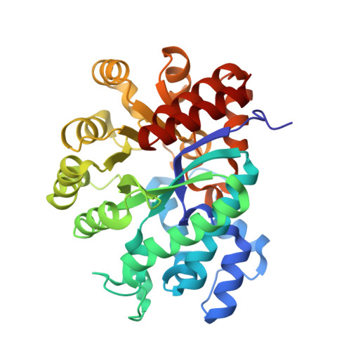
A pre-transition-state mimic of an enzyme: X-ray structure of adenosine deaminase with bound 1-deazaadenosine and zinc-activated water.

Wilson, D.K.Quiocho, F.A.

(1993) Biochemistry 32: 1689-1694

  • DOI: https://doi.org/10.1021/bi00058a001
  • Primary Citation of Related Structures:  
    1ADD

  • PubMed Abstract: 

    The refined 2.4-A structure of adenosine deaminase, recently discovered to be a zinc metalloenzyme [Wilson, D. K., Rudolph, F. B., & Quiocho, F. A. (1991) Science 252, 1278-1284], complexed with the ground-state analog 1-deazaadenosine shows the mode of binding of the analog and, unexpectedly, a zinc-activated water (hydroxide). This structure of a pre-transition-state mimic, combined with that previously determined for the complex with 6(R)-hydroxy-1,6-dihydropurine ribonucleoside, a nearly ideal transition-state analog, sheds new understanding of the precise stereospecificity and hydrolytic catalysis of an important and well-characterized member of a large group of zinc metalloenzymes. As both of these excellent mimics were generated in the active site, they demonstrate a powerful means of dissecting the course of an enzymatic reaction by direct crystallographic analysis.


  • Organizational Affiliation

    Howard Hughes Medical Institute, Baylor College of Medicine, Houston, Texas 77030.


Macromolecules
Find similar proteins by:  (by identity cutoff)  |  3D Structure
Entity ID: 1
MoleculeChains Sequence LengthOrganismDetailsImage
ADENOSINE DEAMINASE349Mus musculusMutation(s): 0 
EC: 3.5.4.4
UniProt
Find proteins for P03958 (Mus musculus)
Explore P03958 
Go to UniProtKB:  P03958
Entity Groups  
Sequence Clusters30% Identity50% Identity70% Identity90% Identity95% Identity100% Identity
UniProt GroupP03958
Sequence Annotations
Expand
  • Reference Sequence
Small Molecules
Ligands 2 Unique
IDChains Name / Formula / InChI Key2D Diagram3D Interactions
1DA
Query on 1DA

Download Ideal Coordinates CCD File 
C [auth A]1-DEAZA-ADENOSINE
C11 H14 N4 O4
NVUDDRWKCUAERS-PNHWDRBUSA-N
ZN
Query on ZN

Download Ideal Coordinates CCD File 
B [auth A]ZINC ION
Zn
PTFCDOFLOPIGGS-UHFFFAOYSA-N
Binding Affinity Annotations 
IDSourceBinding Affinity
1DA Binding MOAD:  1ADD Ki: 180 (nM) from 1 assay(s)
PDBBind:  1ADD Ki: 180 (nM) from 1 assay(s)
Experimental Data & Validation

Experimental Data

  • Method: X-RAY DIFFRACTION
  • Resolution: 2.40 Å
  • R-Value Work: 0.181 
  • R-Value Observed: 0.181 
  • Space Group: C 1 2 1
Unit Cell:
Length ( Å )Angle ( ˚ )
a = 102.39α = 90
b = 94.28β = 127.05
c = 73.02γ = 90
Software Package:
Software NamePurpose
X-PLORmodel building
X-PLORrefinement
X-PLORphasing

Structure Validation

View Full Validation Report


Entry History 

Deposition Data

Revision History  (Full details and data files)

  • Version 1.0: 1994-01-31
    Type: Initial release
  • Version 1.1: 2008-03-24
    Changes: Version format compliance
  • Version 1.2: 2011-07-13
    Changes: Version format compliance
  • Version 1.3: 2024-02-07
    Changes: Data collection, Database references, Derived calculations, Other