1ZUW

Crystal structure of B.subtilis glutamate racemase (RacE) with D-Glu


Experimental Data Snapshot

  • Method: X-RAY DIFFRACTION
  • Resolution: 1.75 Å
  • R-Value Free: 0.237 
  • R-Value Work: 0.179 

wwPDB Validation   3D Report Full Report


This is version 1.4 of the entry. See complete history


Literature

Substrate-Induced Conformational Changes in Bacillus subtilis Glutamate Racemase and Their Implications for Drug Discovery

Ruzheinikov, S.N.Taal, M.A.Sedelnikova, S.E.Baker, P.J.Rice, D.W.

(2005) Structure 13: 1707-1713

  • DOI: https://doi.org/10.1016/j.str.2005.07.024
  • Primary Citation of Related Structures:  
    1ZUW

  • PubMed Abstract: 

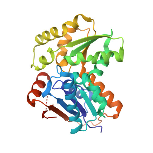
    D-glutamate is an essential building block of the peptidoglycan layer in bacterial cell walls and can be synthesized from L-glutamate by glutamate racemase (RacE). The structure of a complex of B. subtilis RacE with D-glutamate reveals that the glutamate is buried in a deep pocket, whose formation at the interface of the enzyme's two domains involves a large-scale conformational rearrangement. These domains are related by pseudo-2-fold symmetry, which superimposes the two catalytic cysteine residues, which are located at equivalent positions on either side of the alpha carbon of the substrate. The structural similarity of these two domains suggests that the racemase activity of RacE arose as a result of gene duplication. The structure of the complex is dramatically different from that proposed previously and provides new insights into the RacE mechanism and an explanation for the potency of a family of RacE inhibitors, which have been developed as novel antibiotics.


  • Organizational Affiliation

    Krebs Institute for Biomolecular Research, Department of Molecular Biology and Biotechnology, University of Sheffield, Firth Court, Western Bank, Sheffield S10 2TN, United Kingdom.


Macromolecules
Find similar proteins by:  (by identity cutoff)  |  3D Structure
Entity ID: 1
MoleculeChains Sequence LengthOrganismDetailsImage
glutamate racemase 1
A, B, C
272Bacillus subtilisMutation(s): 0 
Gene Names: racE
EC: 5.1.1.3
UniProt
Find proteins for P94556 (Bacillus subtilis (strain 168))
Explore P94556 
Go to UniProtKB:  P94556
Entity Groups  
Sequence Clusters30% Identity50% Identity70% Identity90% Identity95% Identity100% Identity
UniProt GroupP94556
Sequence Annotations
Expand
  • Reference Sequence
Experimental Data & Validation

Experimental Data

  • Method: X-RAY DIFFRACTION
  • Resolution: 1.75 Å
  • R-Value Free: 0.237 
  • R-Value Work: 0.179 
  • Space Group: C 1 2 1
Unit Cell:
Length ( Å )Angle ( ˚ )
a = 134.12α = 90
b = 60.274β = 117.34
c = 125.271γ = 90
Software Package:
Software NamePurpose
DENZOdata reduction
SCALEPACKdata scaling
SHELXSphasing
CNSrefinement

Structure Validation

View Full Validation Report


Entry History 

Deposition Data

Revision History  (Full details and data files)

  • Version 1.0: 2005-12-06
    Type: Initial release
  • Version 1.1: 2008-04-30
    Changes: Version format compliance
  • Version 1.2: 2011-07-13
    Changes: Version format compliance
  • Version 1.3: 2011-11-16
    Changes: Atomic model
  • Version 1.4: 2024-02-14
    Changes: Data collection, Database references, Derived calculations