1ZEN

CLASS II FRUCTOSE-1,6-BISPHOSPHATE ALDOLASE


Experimental Data Snapshot

  • Method: X-RAY DIFFRACTION
  • Resolution: 2.50 Å
  • R-Value Free: 0.326 
  • R-Value Work: 0.243 
  • R-Value Observed: 0.243 

wwPDB Validation   3D Report Full Report


This is version 1.3 of the entry. See complete history


Literature

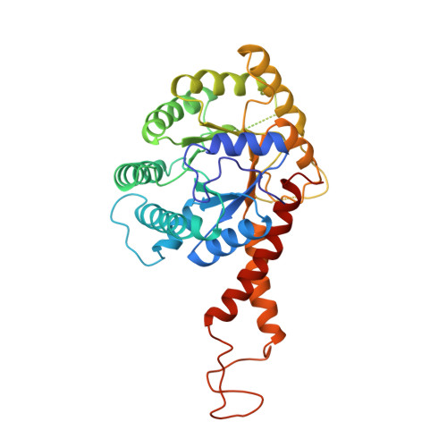
The crystal structure of a class II fructose-1,6-bisphosphate aldolase shows a novel binuclear metal-binding active site embedded in a familiar fold.

Cooper, S.J.Leonard, G.A.McSweeney, S.M.Thompson, A.W.Naismith, J.H.Qamar, S.Plater, A.Berry, A.Hunter, W.N.

(1996) Structure 4: 1303-1315

  • DOI: https://doi.org/10.1016/s0969-2126(96)00138-4
  • Primary Citation of Related Structures:  
    1ZEN

  • PubMed Abstract: 

    [corrected] Aldolases catalyze a variety of condensation and cleavage reactions, with exquisite control on the stereochemistry. These enzymes, therefore, are attractive catalysts for synthetic chemistry. There are two classes of aldolase: class I aldolases utilize Schiff base formation with an active-site lysine whilst class II enzymes require a divalent metal ion, in particular zinc. Fructose-1,6-bisphosphate aldolase (FBP-aldolase) is used in gluconeogenesis and glycolysis; the enzyme controls the condensation of dihydroxyacetone phosphate with glyceraldehyde-3-phosphate to yield fructose-1,6-bisphosphate. Structures are available for class I FBP-aldolases but there is a paucity of detail on the class II enzymes. Characterization is sought to enable a dissection of structure/activity relationships which may assist the construction of designed aldolases for use as biocatalysts in synthetic chemistry.


  • Organizational Affiliation

    Department of Chemistry, University of Manchester, Oxford Road, Manchester, M13 9PL, UK. wnhunter@bad.dundee.ac.uk


Macromolecules
Find similar proteins by:  (by identity cutoff)  |  3D Structure
Entity ID: 1
MoleculeChains Sequence LengthOrganismDetailsImage
CLASS II FRUCTOSE-1,6-BISPHOSPHATE ALDOLASE358Escherichia coliMutation(s): 0 
EC: 4.1.2.13
UniProt
Find proteins for P0AB71 (Escherichia coli (strain K12))
Explore P0AB71 
Go to UniProtKB:  P0AB71
Entity Groups  
Sequence Clusters30% Identity50% Identity70% Identity90% Identity95% Identity100% Identity
UniProt GroupP0AB71
Sequence Annotations
Expand
  • Reference Sequence
Experimental Data & Validation

Experimental Data

  • Method: X-RAY DIFFRACTION
  • Resolution: 2.50 Å
  • R-Value Free: 0.326 
  • R-Value Work: 0.243 
  • R-Value Observed: 0.243 
  • Space Group: P 61 2 2
Unit Cell:
Length ( Å )Angle ( ˚ )
a = 78.36α = 90
b = 78.36β = 90
c = 290.33γ = 120
Software Package:
Software NamePurpose
X-PLORmodel building
X-PLORrefinement
CCP4data reduction
X-PLORphasing

Structure Validation

View Full Validation Report


Entry History 

Deposition Data

Revision History  (Full details and data files)

  • Version 1.0: 1997-07-07
    Type: Initial release
  • Version 1.1: 2008-03-24
    Changes: Version format compliance
  • Version 1.2: 2011-07-13
    Changes: Derived calculations, Version format compliance
  • Version 1.3: 2024-02-14
    Changes: Data collection, Database references, Derived calculations, Other