1YT3

Crystal Structure of Escherichia coli RNase D, an exoribonuclease involved in structured RNA processing


Experimental Data Snapshot

  • Method: X-RAY DIFFRACTION
  • Resolution: 1.60 Å
  • R-Value Free: 0.216 
  • R-Value Work: 0.196 
  • R-Value Observed: 0.196 

wwPDB Validation   3D Report Full Report


This is version 1.4 of the entry. See complete history


Literature

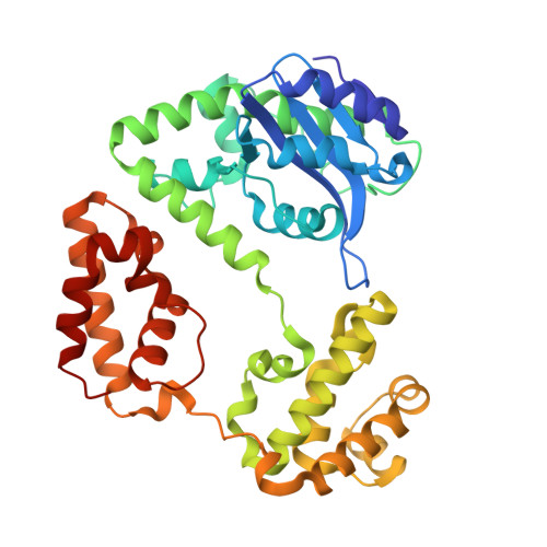
Crystal Structure of Escherichia coli RNase D, an Exoribonuclease Involved in Structured RNA Processing

Zuo, Y.Wang, Y.Malhotra, A.

(2005) Structure 13: 973-984

  • DOI: https://doi.org/10.1016/j.str.2005.04.015
  • Primary Citation of Related Structures:  
    1YT3

  • PubMed Abstract: 

    RNase D (RND) is one of seven exoribonucleases identified in Escherichia coli. RNase D has homologs in many eubacteria and eukaryotes, and has been shown to contribute to the 3' maturation of several stable RNAs. Here, we report the 1.6 A resolution crystal structure of E. coli RNase D. The conserved DEDD residues of RNase D fold into an arrangement very similar to the Klenow fragment exonuclease domain. Besides the catalytic domain, RNase D also contains two structurally similar alpha-helical domains with no discernible sequence homology between them. These closely resemble the HRDC domain previously seen in RecQ-family helicases and several other proteins acting on nucleic acids. More interestingly, the DEDD catalytic domain and the two helical domains come together to form a ring-shaped structure. The ring-shaped architecture of E. coli RNase D and the HRDC domains likely play a major role in determining the substrate specificity of this exoribonuclease.


  • Organizational Affiliation

    Department of Biochemistry and Molecular Biology, University of Miami School of Medicine, PO Box 016129, Miami, FL 33101, USA.


Macromolecules
Find similar proteins by:  (by identity cutoff)  |  3D Structure
Entity ID: 1
MoleculeChains Sequence LengthOrganismDetailsImage
Ribonuclease D375Escherichia coliMutation(s): 0 
Gene Names: RND
EC: 3.1.26.3
UniProt
Find proteins for P09155 (Escherichia coli (strain K12))
Explore P09155 
Go to UniProtKB:  P09155
Entity Groups  
Sequence Clusters30% Identity50% Identity70% Identity90% Identity95% Identity100% Identity
UniProt GroupP09155
Sequence Annotations
Expand
  • Reference Sequence
Small Molecules
Ligands 2 Unique
IDChains Name / Formula / InChI Key2D Diagram3D Interactions
SO4
Query on SO4

Download Ideal Coordinates CCD File 
B [auth A]
C [auth A]
D [auth A]
E [auth A]
F [auth A]
B [auth A],
C [auth A],
D [auth A],
E [auth A],
F [auth A],
G [auth A],
H [auth A]
SULFATE ION
O4 S
QAOWNCQODCNURD-UHFFFAOYSA-L
ZN
Query on ZN

Download Ideal Coordinates CCD File 
I [auth A],
J [auth A],
K [auth A],
L [auth A],
M [auth A]
ZINC ION
Zn
PTFCDOFLOPIGGS-UHFFFAOYSA-N
Experimental Data & Validation

Experimental Data

  • Method: X-RAY DIFFRACTION
  • Resolution: 1.60 Å
  • R-Value Free: 0.216 
  • R-Value Work: 0.196 
  • R-Value Observed: 0.196 
  • Space Group: P 21 21 21
Unit Cell:
Length ( Å )Angle ( ˚ )
a = 51α = 90
b = 80.1β = 90
c = 103.7γ = 90
Software Package:
Software NamePurpose
DENZOdata reduction
SCALEPACKdata scaling
CNSrefinement
CNSphasing

Structure Validation

View Full Validation Report


Entry History 

Deposition Data

Revision History  (Full details and data files)

  • Version 1.0: 2005-08-09
    Type: Initial release
  • Version 1.1: 2008-04-30
    Changes: Version format compliance
  • Version 1.2: 2011-07-13
    Changes: Version format compliance
  • Version 1.3: 2024-02-14
    Changes: Data collection, Database references, Derived calculations
  • Version 1.4: 2024-04-03
    Changes: Refinement description