1Y3I

Crystal Structure of Mycobacterium tuberculosis NAD kinase-NAD complex


Experimental Data Snapshot

  • Method: X-RAY DIFFRACTION
  • Resolution: 2.60 Å
  • R-Value Free: 0.215 

wwPDB Validation   3D Report Full Report


Ligand Structure Quality Assessment 


This is version 1.3 of the entry. See complete history


Literature

NAD-binding mode and the significance of intersubunit contact revealed by the crystal structure of Mycobacterium tuberculosis NAD kinase-NAD complex

Mori, S.Yamasaki, M.Maruyama, Y.Momma, K.Kawai, S.Hashimoto, W.Mikami, B.Murata, K.

(2005) Biochem Biophys Res Commun 327: 500-508

  • DOI: https://doi.org/10.1016/j.bbrc.2004.11.163
  • Primary Citation of Related Structures:  
    1Y3H, 1Y3I

  • PubMed Abstract: 

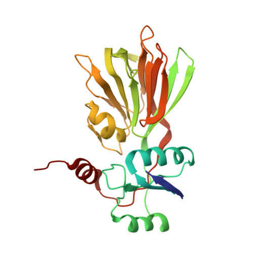
    NAD kinase is a key enzyme in NADP biosynthesis. We solved the crystal structure of polyphosphate/ATP-NAD kinase from Mycobacterium tuberculosis (Ppnk) complexed with NAD (Ppnk-NAD) at 2.6A resolution using apo-Ppnk structure solved in this work, and revealed the details of the structure and NAD-binding site. Superimposition of tertiary structures of apo-Ppnk and Ppnk-NAD demonstrated a substantial conformational difference in a loop (Ppnk-flexible loop). As a quaternary structure, these Ppnk structures exhibited tetramer as in solution condition. Notably, the Ppnk-flexible loop was involved in the intersubunit contact and probably related to the NAD-binding of the other subunit. Furthermore, the two residues (Asp189, His226) substantially contributed to creating NAD-binding site on the other subunit. The two residues and the residues involved in NAD-binding were conserved. However, residues corresponding to the Ppnk-flexible loop were not conserved, making us to speculate that the Ppnk-flexible loop may be Ppnk-specific.


  • Organizational Affiliation

    Department of Basic and Applied Molecular Biotechnology, Graduate School of Agriculture, Kyoto University, Uji, Kyoto 611-0011, Japan.


Macromolecules
Find similar proteins by:  (by identity cutoff)  |  3D Structure
Entity ID: 1
MoleculeChains Sequence LengthOrganismDetailsImage
Inorganic polyphosphate/ATP-NAD kinase
A, B
307Mycobacterium tuberculosisMutation(s): 0 
Gene Names: pPnk
EC: 2.7.1.23
UniProt
Find proteins for P9WHV7 (Mycobacterium tuberculosis (strain ATCC 25618 / H37Rv))
Explore P9WHV7 
Go to UniProtKB:  P9WHV7
Entity Groups  
Sequence Clusters30% Identity50% Identity70% Identity90% Identity95% Identity100% Identity
UniProt GroupP9WHV7
Sequence Annotations
Expand
  • Reference Sequence
Experimental Data & Validation

Experimental Data

  • Method: X-RAY DIFFRACTION
  • Resolution: 2.60 Å
  • R-Value Free: 0.215 
  • Space Group: P 62
Unit Cell:
Length ( Å )Angle ( ˚ )
a = 110.45α = 90
b = 110.45β = 90
c = 108.932γ = 120
Software Package:
Software NamePurpose
SHELXmodel building
SHELXL-97refinement
HKL-2000data reduction
SCALEPACKdata scaling
SHELXphasing

Structure Validation

View Full Validation Report


Ligand Structure Quality Assessment 


Entry History 

Deposition Data

Revision History  (Full details and data files)

  • Version 1.0: 2005-01-18
    Type: Initial release
  • Version 1.1: 2008-04-30
    Changes: Version format compliance
  • Version 1.2: 2011-07-13
    Changes: Derived calculations, Version format compliance
  • Version 1.3: 2024-03-13
    Changes: Data collection, Database references, Derived calculations