1XUQ

Crystal Structure of SodA-1 (BA4499) from Bacillus anthracis at 1.8A Resolution.


Experimental Data Snapshot

  • Method: X-RAY DIFFRACTION
  • Resolution: 1.80 Å
  • R-Value Free: 0.228 
  • R-Value Work: 0.169 
  • R-Value Observed: 0.172 

wwPDB Validation   3D Report Full Report


This is version 1.3 of the entry. See complete history


Literature

Structures of two superoxide dismutases from Bacillus anthracis reveal a novel active centre.

Boucher, I.W.Kalliomaa, A.K.Levdikov, V.M.Blagova, E.V.Fogg, M.J.Brannigan, J.A.Wilson, K.S.Wilkinson, A.J.

(2005) Acta Crystallogr Sect F Struct Biol Cryst Commun 61: 621-624

  • DOI: https://doi.org/10.1107/S1744309105017380
  • Primary Citation of Related Structures:  
    1XRE, 1XUQ

  • PubMed Abstract: 

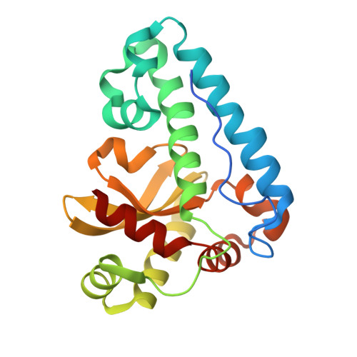
    The BA4499 and BA5696 genes of Bacillus anthracis encode proteins homologous to manganese superoxide dismutase, suggesting that this organism has an expanded repertoire of antioxidant proteins. Differences in metal specificity and quaternary structure between the dismutases of prokaryotes and higher eukaryotes may be exploited in the development of therapeutic antibacterial compounds. Here, the crystal structure of two Mn superoxide dismutases from B. anthracis solved to high resolution are reported. Comparison of their structures reveals that a highly conserved residue near the active centre is substituted in one of the proteins and that this is a characteristic feature of superoxide dismutases from the B. cereus/B. anthracis/B. thuringiensis group of organisms.


  • Organizational Affiliation

    Department of Chemistry, University of York, York YO10 5YW, England.


Macromolecules
Find similar proteins by:  (by identity cutoff)  |  3D Structure
Entity ID: 1
MoleculeChains Sequence LengthOrganismDetailsImage
Superoxide dismutase
A, B
212Bacillus anthracisMutation(s): 0 
Gene Names: sodA1sodA-1
EC: 1.15.1.1
UniProt
Find proteins for Q81LW0 (Bacillus anthracis)
Explore Q81LW0 
Go to UniProtKB:  Q81LW0
Entity Groups  
Sequence Clusters30% Identity50% Identity70% Identity90% Identity95% Identity100% Identity
UniProt GroupQ81LW0
Sequence Annotations
Expand
  • Reference Sequence
Experimental Data & Validation

Experimental Data

  • Method: X-RAY DIFFRACTION
  • Resolution: 1.80 Å
  • R-Value Free: 0.228 
  • R-Value Work: 0.169 
  • R-Value Observed: 0.172 
  • Space Group: P 1 21 1
Unit Cell:
Length ( Å )Angle ( ˚ )
a = 60.881α = 90
b = 60.439β = 106.3
c = 61.981γ = 90
Software Package:
Software NamePurpose
REFMACrefinement
HKL-2000data reduction
SCALEPACKdata scaling
MOLREPphasing

Structure Validation

View Full Validation Report


Entry History 

Deposition Data

Revision History  (Full details and data files)

  • Version 1.0: 2005-07-19
    Type: Initial release
  • Version 1.1: 2007-10-16
    Changes: Version format compliance
  • Version 1.2: 2011-07-13
    Changes: Version format compliance
  • Version 1.3: 2023-08-23
    Changes: Data collection, Database references, Derived calculations, Refinement description