1XOF

Heterooligomeric Beta Beta Alpha Miniprotein


Experimental Data Snapshot

  • Method: X-RAY DIFFRACTION
  • Resolution: 1.95 Å
  • R-Value Free: 0.240 
  • R-Value Work: 0.222 
  • R-Value Observed: 0.222 

wwPDB Validation   3D Report Full Report


This is version 2.0 of the entry. See complete history


Literature

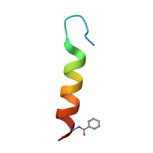
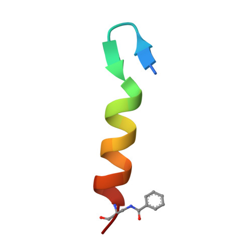
Design of a Heterospecific, Tetrameric, 21-Residue Miniprotein with Mixed alpha/beta Structure.

Ali, M.H.Taylor, C.M.Grigoryan, G.Allen, K.N.Imperiali, B.Keating, A.E.

(2005) Structure 13: 225-234

  • DOI: https://doi.org/10.1016/j.str.2004.12.009
  • Primary Citation of Related Structures:  
    1XOF

  • PubMed Abstract: 

    The study of short, autonomously folding peptides, or "miniproteins," is important for advancing our understanding of protein stability and folding specificity. Although many examples of synthetic alpha-helical structures are known, relatively few mixed alpha/beta structures have been successfully designed. Only one mixed-secondary structure oligomer, an alpha/beta homotetramer, has been reported thus far. In this report, we use structural analysis and computational design to convert this homotetramer into the smallest known alpha/beta-heterotetramer. Computational screening of many possible sequence/structure combinations led efficiently to the design of short, 21-residue peptides that fold cooperatively and autonomously into a specific complex in solution. A 1.95 A crystal structure reveals how steric complementarity and charge patterning encode heterospecificity. The first- and second-generation heterotetrameric miniproteins described here will be useful as simple models for the analysis of protein-protein interaction specificity and as structural platforms for the further elaboration of folding and function.


  • Organizational Affiliation

    Department of Chemistry, Massachusetts Institute of Technology, 77 Massachusetts Avenue, Cambridge, Massachusetts 02139, USA.


Macromolecules

Find similar proteins by:  Sequence   |   3D Structure  

Entity ID: 1
MoleculeChains Sequence LengthOrganismDetailsImage
BBAhetT122N/AMutation(s): 0 
Entity Groups  
Sequence Clusters30% Identity50% Identity70% Identity90% Identity95% Identity100% Identity
Sequence Annotations
Expand
  • Reference Sequence

Find similar proteins by:  Sequence   |   3D Structure  

Entity ID: 2
MoleculeChains Sequence LengthOrganismDetailsImage
BBAhetT122N/AMutation(s): 0 
Entity Groups  
Sequence Clusters30% Identity50% Identity70% Identity90% Identity95% Identity100% Identity
Sequence Annotations
Expand
  • Reference Sequence
Small Molecules
Modified Residues  1 Unique
IDChains TypeFormula2D DiagramParent
DBZ
Query on DBZ
A
L-PEPTIDE LINKINGC10 H12 N2 O3ALA
Experimental Data & Validation

Experimental Data

  • Method: X-RAY DIFFRACTION
  • Resolution: 1.95 Å
  • R-Value Free: 0.240 
  • R-Value Work: 0.222 
  • R-Value Observed: 0.222 
  • Space Group: P 31 2 1
Unit Cell:
Length ( Å )Angle ( ˚ )
a = 41.695α = 90
b = 41.695β = 90
c = 51.332γ = 120
Software Package:
Software NamePurpose
CNSrefinement
HKL-2000data reduction
SCALEPACKdata scaling
MOLREPphasing

Structure Validation

View Full Validation Report


Entry History 

Deposition Data

Revision History  (Full details and data files)

  • Version 1.0: 2005-02-01
    Type: Initial release
  • Version 1.1: 2008-04-30
    Changes: Version format compliance
  • Version 1.2: 2011-07-13
    Changes: Version format compliance
  • Version 1.3: 2018-01-31
    Changes: Experimental preparation
  • Version 2.0: 2023-11-15
    Changes: Atomic model, Data collection, Database references, Derived calculations