Structures of the DNA-binding site of Runt-domain transcription regulators.
Kitayner, M., Rozenberg, H., Rabinovich, D., Shakked, Z.(2005) Acta Crystallogr D Biol Crystallogr 61: 236-246
- PubMed: 15735333
- DOI: https://doi.org/10.1107/S0907444904032378
- Primary Citation of Related Structures:
1XJX, 1XJY - PubMed Abstract:
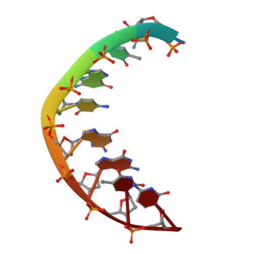
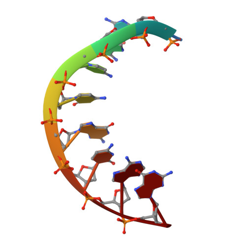
Runt-domain (RD) proteins are transcription factors that play fundamental roles in various developmental pathways. They bind specifically to DNA sequences of the general form PyGPyGGTPy (Py = pyrimidine), through which they regulate transcription of target genes. The DNA duplex TCTGCGGTC/TGACCGCAG, incorporating the binding site for the RD transcription factors (bold), was crystallized in space group P4(3). X-ray analysis of two crystals diffracting to 1.7 and 2.0 angstroms resolution, which had slight variations in their unit-cell parameters, revealed two distinct conformations of the A-DNA helix. The two crystal structures possessed several structure and hydration features that had previously been observed in A-DNA duplexes. A comparative analysis of the present A-DNA structures and those of previously reported B-DNA crystal structures of RD-binding sites in free and protein-bound states showed the various duplexes to display several common features. Within this series, the present A-DNA duplexes adopt two conformations along the pathway from the canonical A-DNA to the B-DNA forms and the protein-bound helices display conformational features that are intermediate between those of the current A-DNA structures and that of the B-DNA-type helix of the free RD target. Based on these data and energy considerations, it is likely that the propensity of the RD-binding site to adopt the A-DNA or B-DNA conformation in solution depends on the sequence context and environmental conditions, and that the transition from either DNA form to the protein-bound conformation involves a small energy barrier.
Organizational Affiliation:
Department of Structural Biology, Weizmann Institute of Science, Rehovot 76100, Israel.