1X04

Crystal structure of endophilin BAR domain (mutant)


Experimental Data Snapshot

  • Method: X-RAY DIFFRACTION
  • Resolution: 2.90 Å
  • R-Value Free: 0.269 
  • R-Value Work: 0.238 
  • R-Value Observed: 0.240 

wwPDB Validation   3D Report Full Report


This is version 1.3 of the entry. See complete history


Literature

Endophilin BAR domain drives membrane curvature by two newly identified structure-based mechanisms

Masuda, M.Takeda, S.Sone, M.Ohki, T.Mori, H.Kamioka, Y.Mochizuki, N.

(2006) EMBO J 25: 2889-2897

  • DOI: https://doi.org/10.1038/sj.emboj.7601176
  • Primary Citation of Related Structures:  
    1X03, 1X04, 2D4C

  • PubMed Abstract: 

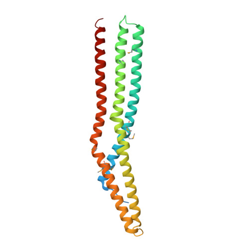
    The crescent-shaped BAR (Bin/Amphiphysin/Rvs-homology) domain dimer is a versatile protein module that senses and generates positive membrane curvature. The BAR domain dimer of human endophilin-A1, solved at 3.1 A, has a unique structure consisting of a pair of helix-loop appendages sprouting out from the crescent. The appendage's short helices form a hydrophobic ridge, which runs across the concave surface at its center. Examining liposome binding and tubulation in vitro using purified BAR domain and its mutants indicated that the ridge penetrates into the membrane bilayer and enhances liposome tubulation. BAR domain-expressing cells exhibited marked plasma membrane tubulation in vivo. Furthermore, a swinging-arm mutant lost liposome tubulation activity yet retaining liposome binding. These data suggested that the rigid crescent dimer shape is crucial for the tubulation. We here propose that the BAR domain drives membrane curvature by coordinate action of the crescent's scaffold mechanism and the ridge's membrane insertion in addition to membrane binding via amino-terminal amphipathic helix.


  • Organizational Affiliation

    Department of Structural Analysis, National Cardiovascular Center Research Institute, Suita, Osaka, Japan.


Macromolecules
Find similar proteins by:  (by identity cutoff)  |  3D Structure
Entity ID: 1
MoleculeChains Sequence LengthOrganismDetailsImage
SH3-containing GRB2-like protein 2230Homo sapiensMutation(s): 8 
EC: 2.3.1
UniProt & NIH Common Fund Data Resources
Find proteins for Q99962 (Homo sapiens)
Explore Q99962 
Go to UniProtKB:  Q99962
PHAROS:  Q99962
GTEx:  ENSG00000107295 
Entity Groups  
Sequence Clusters30% Identity50% Identity70% Identity90% Identity95% Identity100% Identity
UniProt GroupQ99962
Sequence Annotations
Expand
  • Reference Sequence
Small Molecules
Modified Residues  1 Unique
IDChains TypeFormula2D DiagramParent
MSE
Query on MSE
A
L-PEPTIDE LINKINGC5 H11 N O2 SeMET
Experimental Data & Validation

Experimental Data

  • Method: X-RAY DIFFRACTION
  • Resolution: 2.90 Å
  • R-Value Free: 0.269 
  • R-Value Work: 0.238 
  • R-Value Observed: 0.240 
  • Space Group: I 41
Unit Cell:
Length ( Å )Angle ( ˚ )
a = 129.077α = 90
b = 129.077β = 90
c = 97.963γ = 90
Software Package:
Software NamePurpose
HKL-2000data collection
SCALEPACKdata scaling
MOLREPphasing
CNSrefinement
HKL-2000data reduction

Structure Validation

View Full Validation Report


Entry History 

Deposition Data

Revision History  (Full details and data files)

  • Version 1.0: 2006-05-02
    Type: Initial release
  • Version 1.1: 2008-04-30
    Changes: Version format compliance
  • Version 1.2: 2011-07-13
    Changes: Derived calculations, Version format compliance
  • Version 1.3: 2017-08-23
    Changes: Refinement description, Source and taxonomy