1WZZ

Structure of endo-beta-1,4-glucanase CMCax from Acetobacter xylinum


Experimental Data Snapshot

  • Method: X-RAY DIFFRACTION
  • Resolution: 1.65 Å
  • R-Value Free: 0.195 
  • R-Value Work: 0.176 

wwPDB Validation   3D Report Full Report


This is version 1.2 of the entry. See complete history


Literature

Structural characterization of the Acetobacter xylinum endo-beta-1,4-glucanase CMCax required for cellulose biosynthesis.

Yasutake, Y.Kawano, S.Tajima, K.Yao, M.Satoh, Y.Munekata, M.Tanaka, I.

(2006) Proteins 64: 1069-1077

  • DOI: https://doi.org/10.1002/prot.21052
  • Primary Citation of Related Structures:  
    1WZZ

  • PubMed Abstract: 

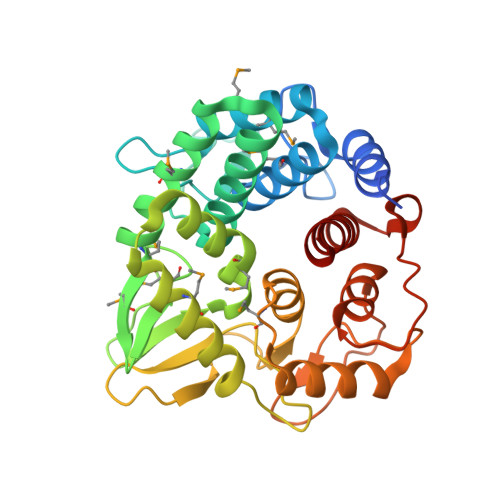
    Previous studies have demonstrated that endoglucanase is required for cellulose biosynthesis both in bacteria and plants. However, it has yet to be elucidated how the endoglucanases function in the mechanism of cellulose biosynthesis. Here we describe the crystal structure of the cellulose biosynthesis-related endo-beta-1,47-glucanase (CMCax; EC 3.2.1.4) from the cellulose-producing Gramnegative bacterium, Acetobacter xylinum (= Gluconacetobacter xylinus), determined at 1.65-A resolution. CMCax falls into the glycoside hydrolase family 8 (GH-8), and the structure showed that the overall fold of the CMCax is similar to those of other glycoside hydrolases belonging to GH-8. Structure comparison with Clostridium thermocellum CelA, the best characterized GH-8 endoglucanase, revealed that sugar recognition subsite +3 is completely missing in CMCax. The absence of the subsite +3 leads to significant broadness of the cleft at the cellooligosaccharide reducing-end side. CMCax is known to be a secreted enzyme and is present in the culture medium. However, electron microscopic analysis using immunostaining clearly demonstrated that a portion of CMCax is localized to the cell surface, suggesting a link with other known membrane-anchored endoglucanases that are required for cellulose biosynthesis.


  • Organizational Affiliation

    Division of Biological Sciences, Graduate School of Science, Hokkaido University, Sapporo, Hokkaido 060-0810, Japan.


Macromolecules
Find similar proteins by:  (by identity cutoff)  |  3D Structure
Entity ID: 1
MoleculeChains Sequence LengthOrganismDetailsImage
Probable endoglucanase334Komagataeibacter xylinusMutation(s): 10 
Gene Names: cmcAX
EC: 3.2.1.4
UniProt
Find proteins for P37696 (Novacetimonas hansenii)
Explore P37696 
Go to UniProtKB:  P37696
Entity Groups  
Sequence Clusters30% Identity50% Identity70% Identity90% Identity95% Identity100% Identity
UniProt GroupP37696
Sequence Annotations
Expand
  • Reference Sequence
Small Molecules
Ligands 1 Unique
IDChains Name / Formula / InChI Key2D Diagram3D Interactions
SO4
Query on SO4

Download Ideal Coordinates CCD File 
B [auth A]SULFATE ION
O4 S
QAOWNCQODCNURD-UHFFFAOYSA-L
Modified Residues  1 Unique
IDChains TypeFormula2D DiagramParent
MSE
Query on MSE
A
L-PEPTIDE LINKINGC5 H11 N O2 SeMET
Experimental Data & Validation

Experimental Data

  • Method: X-RAY DIFFRACTION
  • Resolution: 1.65 Å
  • R-Value Free: 0.195 
  • R-Value Work: 0.176 
  • Space Group: P 61
Unit Cell:
Length ( Å )Angle ( ˚ )
a = 88.878α = 90
b = 88.878β = 90
c = 93.98γ = 120
Software Package:
Software NamePurpose
DENZOdata reduction
SCALEPACKdata scaling
SHELXDEphasing
CNSrefinement
SHELXDphasing
SHELXEmodel building

Structure Validation

View Full Validation Report


Entry History 

Deposition Data

Revision History  (Full details and data files)

  • Version 1.0: 2006-03-14
    Type: Initial release
  • Version 1.1: 2008-04-30
    Changes: Version format compliance
  • Version 1.2: 2011-07-13
    Changes: Version format compliance