1WVF

p-Cresol Methylhydroxylase: Alteration of the Structure of the Flavoprotein Subunit upon its Binding to the Cytochrome Subunit


Experimental Data Snapshot

  • Method: X-RAY DIFFRACTION
  • Resolution: 1.30 Å
  • R-Value Free: 0.167 
  • R-Value Work: 0.146 

wwPDB Validation   3D Report Full Report


Ligand Structure Quality Assessment 


This is version 1.3 of the entry. See complete history


Literature

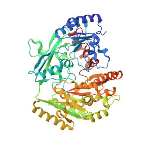
p-Cresol Methylhydroxylase: Alteration of the Structure of the Flavoprotein Subunit upon Its Binding to the Cytochrome Subunit

Cunane, L.M.Chen, Z.-W.McIntire, W.S.Mathews, F.S.

(2005) Biochemistry 44: 2963-2973

  • DOI: https://doi.org/10.1021/bi048020r
  • Primary Citation of Related Structures:  
    1WVE, 1WVF

  • PubMed Abstract: 

    The structures of two forms of a recombinant flavoprotein have been determined at high resolution and compared. These proteins are (1) the flavocytochrome c p-cresol methylhydroxylase (rPCMH, 1.85 A resolution) and (2) the cytochrome-free flavoprotein subunit of rPCMH (PchF, 1.30 A resolution). A significant conformational difference is observed in a protein segment that is in contact with the re face of the isoalloxazine ring of FAD when the structure of PchF is compared to the subunit in the intact flavocytochrome. This structural change is important for optimum catalytic function of the flavoprotein, which has been shown to be dependent on the presence of the cytochrome subunit. This change results in different protein-flavin and apparently different protein-substrate interactions that have a "tuning effect" on the electronic and redox properties of bound p-cresol and the covalently bound FAD. The conformational change in the segment in the cofactor-binding site is induced by a small rearrangement in the flavoprotein-cytochrome interface region of the flavoprotein.


  • Organizational Affiliation

    Department of Biochemistry and Molecular Biophysics, Washington University School of Medicine, St. Louis, Missouri 63110, USA.


Macromolecules
Find similar proteins by:  (by identity cutoff)  |  3D Structure
Entity ID: 1
MoleculeChains Sequence LengthOrganismDetailsImage
4-cresol dehydrogenase [hydroxylating] flavoprotein subunit520Pseudomonas putidaMutation(s): 0 
EC: 1.17.99.1
UniProt
Find proteins for P09788 (Pseudomonas putida)
Explore P09788 
Go to UniProtKB:  P09788
Entity Groups  
Sequence Clusters30% Identity50% Identity70% Identity90% Identity95% Identity100% Identity
UniProt GroupP09788
Sequence Annotations
Expand
  • Reference Sequence
Experimental Data & Validation

Experimental Data

  • Method: X-RAY DIFFRACTION
  • Resolution: 1.30 Å
  • R-Value Free: 0.167 
  • R-Value Work: 0.146 
  • Space Group: C 1 2 1
Unit Cell:
Length ( Å )Angle ( ˚ )
a = 88.64α = 90
b = 116.91β = 113.18
c = 50.22γ = 90
Software Package:
Software NamePurpose
DENZOdata reduction
SCALEPACKdata scaling
AMoREphasing
CNSrefinement

Structure Validation

View Full Validation Report


Ligand Structure Quality Assessment 


Entry History 

Deposition Data

Revision History  (Full details and data files)

  • Version 1.0: 2005-03-08
    Type: Initial release
  • Version 1.1: 2008-04-30
    Changes: Version format compliance
  • Version 1.2: 2011-07-13
    Changes: Derived calculations, Version format compliance
  • Version 1.3: 2023-10-25
    Changes: Data collection, Database references, Derived calculations, Refinement description